Download
1 / 1
20 likes | 567 Vues
Improved growth of Vancomycin resistant Enterococci on ChromID™ VRE agar by incubation in 5% CO 2. Varuna Navaratne, Sydney Bell and Jeanette Pham SEALS Microbiology, Randwick and Kogarah, NSW. Introduction

E N D
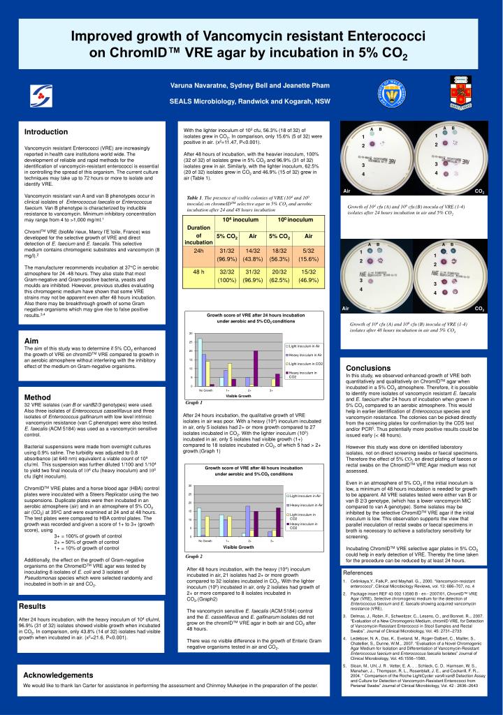
Improved growth of Vancomycin resistant Enterococci on ChromID™ VRE agar by incubation in 5% CO2 Varuna Navaratne, Sydney Bell and Jeanette Pham SEALS Microbiology, Randwick and Kogarah, NSW Introduction Vancomycin resistant Enterococci (VRE) are increasingly reported in health care institutions world wide. The development of reliable and rapid methods for the identification of vancomycin-resistant enterococci is essential in controlling the spread of this organism. The current culture techniques may take up to 72 hours or more to isolate and identify VRE. Vancomycin resistant van A and van B phenotypes occur in clinical isolates of Enterococcus faecalis or Enterococcus faecium. Van B phenotype is characterized by inducible resistance to vancomycin. Minimum inhibitory concentration may range from 4 to >1,000 mg/ml.1 ChromITM VRE (bioMe´rieux, Marcy l’E´toile, France) was developed for the selective growth of VRE and direct detection of E. faecium and E. faecalis. This selective medium contains chromogenic substrates and vancomycin (8 mg/l).2 The manufacturer recommends incubation at 37°C in aerobic atmosphere for 24 -48 hours. They also state that most Gram-negative and Gram-positive bacteria, yeasts and moulds are inhibited. However, previous studies evaluating this chromogenic medium have shown that some VRE strains may not be apparent even after 48 hours incubation. Also there may be breakthrough growth of some Gram negative organisms which may give rise to false positive results.3,4 With the lighter inoculum of 100 cfu, 56.3% (18 of 32) of isolates grew in CO2. In comparison, only 15.6% (5 of 32) were positive in air. (x2=11.47, P<0.001). After 48 hours of incubation, with the heavier inoculum, 100% (32 of 32) of isolates grew in 5% CO2 and 96.9% (31 of 32) isolates grew in air. Similarly, with the lighter inoculum, 62.5% (20 of 32) isolates grew in CO2 and 46.9% (15 of 32) grew in air (Table 1). A B A B 1 2 3 4 1 2 3 4 Air CO2 Table 1. The presence of visible colonies of VRE (104 and 100 inocula) on chromeIDTM selective agar in 5% CO2 and aerobic incubation after 24 and 48 hours incubation Growth of 104 cfu (A) and 100 cfu (B) inocula of VRE (1-4)isolates after 24 hours incubation in air and 5% CO2 A B A B 1 2 3 4 1 2 3 4 Air CO2 Growth of 104 cfu (A) and 100 cfu (B) inocula of VRE (1-4)isolates after 48 hours incubation in air and 5% CO2 Aim The aim of this study was to determine if 5% CO2 enhanced the growth of VRE on chromIDTM VRE compared to growth in an aerobic atmosphere without interfering with the inhibitory effect of the medium on Gram-negative organisms. Conclusions In this study, we observed enhanced growth of VRE both quantitatively and qualitatively on ChromIDTM agar when incubated in a 5% CO2 atmosphere. Therefore, it is possible to identify more isolates of vancomycin resistant E. faecalis and E. faecium after 24 hours of incubation when grown in 5% CO2 compared to an aerobic atmosphere. This would help in earlier identification of Enterococcus species and vancomycin resistance. The colonies can be picked directly from the screening plates for confirmation by the CDS test and/or PCR5. Thus potentially more positive results could be issued early (< 48 hours). However this study was done on identified laboratory isolates, not on direct screening swabs or faecal specimens. Therefore the effect of 5% CO2 on direct plating of faeces or rectal swabs on the ChromIDTM VRE Agar medium was not assessed. Even in an atmosphere of 5% CO2 if the initial inoculum is low, a minimum of 48 hours incubation is needed for growth to be apparent. All VRE isolates tested were either van B or van B 2/3 genotype, (which has a lower vancomycin MIC compared to van A genotype). Some isolates may be inhibited by the selective ChromIDTM VRE agar if the initial inoculum is low. This observation supports the view that parallel inoculation of rectal swabs or faecal specimens in broth is necessary to achieve a satisfactory sensitivity for screening. Incubating ChromIDTM VRE selective agar plates in 5% CO2 could help in early detection of VRE. Thereby the time taken for the procedure can be reduced by at least 24 hours. • Method • 32 VRE isolates (van B or vanB2/3 genotypes) were used. • Also three isolates of Enterococcus casseliflavus and three • isolates of Enterococcus gallinarum with low level intrinsic • vancomycin resistance (van C phenotype) were also tested. E. faecalis (ACM 5184) was used as a vancomycin sensitive control. • Bacterial suspensions were made from overnight cultures • using 0.9% saline. The turbidity was adjusted to 0.8 • absorbance (at 640 nm) equivalent a viable count of 109 • cfu/ml. This suspension was further diluted 1/100 and 1/104 • to yield two final inocula of 104 cfu (heavy inoculum) and 100 cfu (light inoculum). • ChromIDTM VRE plates and a horse blood agar (HBA) control • plates were inoculated with a Steers Replicator using the two • suspensions. Duplicate plates were then incubated in an • aerobic atmosphere (air) and in an atmosphere of 5% CO2 • air (CO2) at 35oC and were examined at 24 and at 48 hours. The test plates were compared to HBA control plates. The growth was recorded and given a score of 1+ to 3+ (growth score), using • 3+ = 100% of growth of control • 2+ = 50% of growth of control • 1+ = 10% of growth of control • Additionally, the effect on the growth of Gram-negative • organisms on the ChromeIDTM VRE agar was tested by • inoculating 8 isolates of E. coli and 3 isolates of • Pseudomonas species which were selected randomly and • incubated in both in air and CO2. Graph 1 After 24 hours incubation, the qualitative growth of VRE isolates in air was poor. With a heavy (104) inoculum incubated in air, only 5 isolates had 2+ or more growth compared to 27 isolates incubated in CO2. With the lighter inoculum (100) incubated in air, only 5 isolates had visible growth (1+) compared to 18 isolates incubated in CO2, of which 5 had > 2+ growth.(Graph 1) Graph 2 After 48 hours incubation, with the heavy (104) inoculum incubated in air,21 isolates had 2+ or more growth compared to 32 isolates incubated in CO2. With the lighter inoculum (100) incubated in air, only 2 isolates had growth of 2+ or more compared to 8 isolates incubated in CO2.(Graph2) The vancomycin sensitive E. faecalis (ACM 5184) control and the E. casseliflavus and E. gallinarum isolates did not grow on the chromIDTM VRE agar in both air and CO2 after 48 hours. There was no visible difference in the growth of Enteric Gram negative organisms tested in air and CO2. • References • Cetinkaya,Y., Falk,P., and Mayhall. G.,. 2000. “Vancomycin-resistant enterococci”, Clinical Microbiology Reviews, vol. 13: 686–707, no. 4 • Package insert REF 43 002 13580 B - en - 2007/01, ChromID™ VRE Agar (VRE), Selective chromogenic medium for the detection of Enterococcus faecium and E. faecalis showing acquired vancomycin resistance (VRE). • Delmas, J., Robin, F., Schweitzer, C., Lesens, O., and Bonnet. R.,. 2007. “Evaluation of a New Chromogenic Medium, chromID VRE, for Detection of Vancomycin-Resistant Enterococci in Stool Samples and Rectal Swabs”. Journal of Clinical Microbiology, Vol. 45: 2731–2733 • Ledeboer, N. A., Das, K., Eveland, M., Roger-Dalbert, C., Mailler, S., Chatellier, S., Dunne, W.M.,. 2007. “Evaluation of a Novel Chromogenic Agar Medium for Isolation and Differentiation of Vancomycin-Resistant Enterococcus faecium and Enterococcus faecalis Isolates” Journal of Clinical Microbiology, Vol. 45:1556–1560, • Sloan, M.. Uhl, J. R . Vetter, E. A. , . Schleck, C. D, Harmsen, W. S., Manahan, J., Thompson, R. L., Rosenblatt, J. E., and Cockerill, F. R.,. 2004. “ Comparison of the Roche LightCycler vanA/vanB Detection Assay and Culture for Detection of Vancomycin-Resistant Enterococci from Perianal Swabs” Journal of Clinical Microbiology, Vol. 42 : 2636–2643 Results After 24 hours incubation, with the heavy inoculum of 104 cfu/ml,96.9% (31 of 32) isolates showed visible growth when incubated in CO2. In comparison, only 43.8% (14 of 32) isolates had visible growth when incubated in air. (x2=21.6, P<0.001). Acknowledgements We would like to thank Ian Carter for assistance in performing the assessment and Chinmoy Mukerjee in the preparation of the poster.