Download
1 / 42
530 likes | 984 Vues
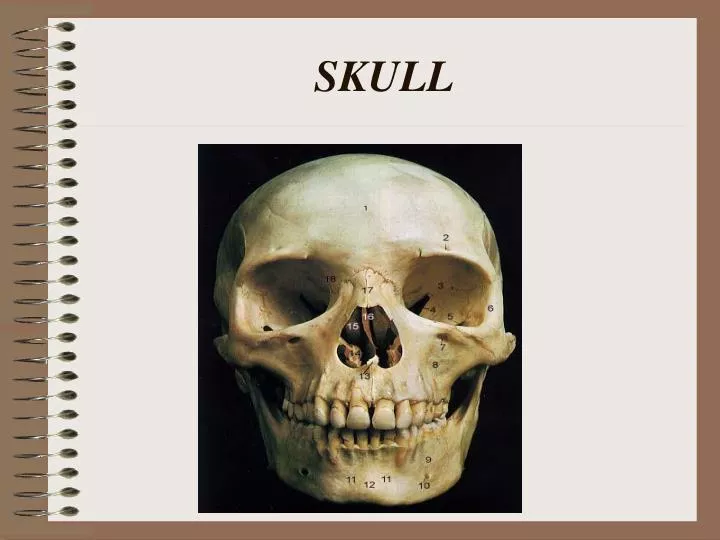
SKULL. HUMAN SKULL. Consists of 22 bones 8 of these bones make up the cranium 14 form the facial skeleton. (also 6 tiny bones in the middle ear). The cranium encloses and protect the brain, and its surface provides attachments for muscles that make chewing and head movements possible.

E N D
HUMAN SKULL • Consists of 22 bones • 8 of these bones make up the cranium • 14 form the facial skeleton. • (also 6 tiny bones in the middle ear)
The cranium encloses and protect the brain, and its surface provides attachments for muscles that make chewing and head movements possible. There are 8 bones that make up the cranium. Frontal Bone Parietal Bones (2) Occipital Bone Temporal Bones (2) Sphenoid Bone Ethmoid Bone PARTS OF THE SKULLCRANIUM
FRONTAL BONE • Forms the anterior portion of the skull above the eyes, including the forehead, the roof of the nasal cavity and roof of the orbits of the eyes.
Parietal Bones The parietal bones (2) form, by their union, the sides and roof of the cranium. Each bone is irregularly quadrilateral in form, and has two surfaces, four borders, and four angles.
Occipital Bone • The occipital bone joins the parietal bones. • It forms the back of the skull and the base of the cranium. • A large opening on its lower surface houses nerve fibers from the brain that pass to the spinal cord.
Temporal Bones • A Temporal bones (2) on each side of the skull joins the parietal bone along a squamosal suture. • Forms the sides and the base of the cranium. • House the internal ear. • Notice how far it goes around the face before it meets the zygomatic bone “cheek”
Sphenoid Bones • The sphenoid bone is wedged between several other bones in the anterior portion of the cranium. • Helps form the base of the cranium, the sides of the skull and the floors and sides of the orbits. • This bone really has three different views… Laterial—anterior to the temporal bone • Anterior- the back of the eye sockets • Inferior- looks like a butterfly.
Sphenoid Bones • This bone really has three different views… • Lateral—anterior to the temporal bone • Anterior- the back of the eye sockets • Inferior- looks like a butterfly.
Ethmoid Bone • Located in front of the sphenoid bone. • Forms part of roof and walls of nasal cavity. • Floor of cranium, and walls of orbits. • In the back of the orbital cavity…longer bone
The facial skeleton consists of 13 immovable bones and a movable lower jaw bone. These bones provide basic shape of the face Attachments for muscles that move the jaw and control facial expressions. Facial Skeleton
MANDIBLE • Lower jaw bone • A moveable bone held to the cranium by ligaments. • Horseshoe-shaped body with a flat ramus projecting upward at each end
MAXILLARY BONES • They form the upper jaw. • Together the form the keystone of the face. • They form anterior roof of mouth, floors of orbits, and floor of nasal cavity. • Contains aleveolar process, maxillary sinuses, palatine process.
The L-shaped palatine bones are located behind the maxillae. The horizontal portions form the posterior section of the hard palate and the floor of the nasal cavity. The perpendicular portions help form the lateral walls of the nasal cavity. This is easier too see from the bottom view of the skull. Palatine Bones
ZYGOMATIC BONES • Responsible for the prominences of the cheeks below and to the sides of the eyes. • Help form the lateral walls and the floors of the orbits. • Again notice where it meets the temporal bone…called the Zygomatic Arch or the Zygomatic process of the Temporal
A thin, scalelike structure located in the medial wall of each orbit between the ethmoid bone and the maxilla. A groove in its anterior portion leads from the orbit to the nasal cavity, providing a pathway for a channel that carries tears from the eye to the nasal cavity. Lacrimal Bones
NASAL BONES • The nasal bones are long, thin, and nearly rectangular. • They lie side by side and are fused at the midline, where they form the bridge of the nose. • These bones are attachments for the cartilaginous tissue that forms the shape of the nose.
The thin, flat vomer bone is located along the midline within the nasal cavity. Posteriorly, it joins the perpendicular plate of the ethmoid bone Together they form the nasal septon. Vomer bone
Inferior Nasal Cavity • The inferior nasal conchae are fragile, scroll-shaped bones attached to the lateral walls of the nasal cavity. • Positioned below the superior and middle nasal conchae of the ethmoid bone.
Foramen • Foramen- An opening through a bone that usually serves as a passageway for blood vessels, nerves, or ligaments. • You need to be able to identify the following… • Mental Foramen, Supraorbital Foramen • Infraorbital Foramen, Foramen Magnum
Foramen Supraorbital foramen (notch) Infraorbital foramen Mental foramen
Foramen Foramen magnum
Sutures • Suture- an interlocking line of union between bones • You need to be familiar with the following sutures…. • Coronal Suture, • Lambdoidal Suture • Squamosal Suture • Sagittal Suture
Sutures Coronal suture Squamous suture Lambdoid suture
Sutures Sagittal suture Lambdoid suture
Process • Process- a prominent projection on a bone • You need to familiar with the following… • Styloid Process • Mastoid Process • Coronoid Process
Process Mastoid process Styloid process Coronoid process
Condyle • Condyle- is a rounded process that usually articulates with another bone • You need to be familiar with the following… • Mandibular Condyle • Occipital Condyle
Condyle Mandibular condyle
Condyle Occipital condyle
Inferior View Palatine bone Zygomaticbone Sphenoid bone Vomer
For Your Information Zygomatic process (Aka zygomatic arch External acoustic canal
Ear Bones • Malleus (2)- means hammer- shape of the bone • Incus(2)- means anvil- shape of bone • Stapes (2) means stirrup- shape of the bone • Don’t worry about identification of these on the skull…you cannot see them. Just recognize the names as bones of the inner ear.
What have we learned so far?? Coronal Suture FrontalBone SphenoidBone ParietalBone Ethmoid Bone Temporal Bone Lacrimal Bones Lamboid suture Squamous suture Nasal Bone Occipital bone ZygomaticBone Maxilla External Auditory Canal Mastoid Process Mandible Styloid Process Mandibular condyle Mental Foramen Coronoid process