Download

1 / 32
490 likes | 2.82k Vues
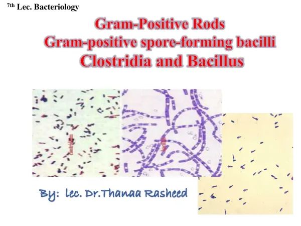
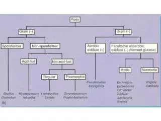
Aerobic Non-Spore Forming Gram-Positive Bacilli. Corynebacterium. Gram Positive Bacilli. Gram positive rods. Spore forming. Non spore forming. Aerobic. Anaerobic. Corynebacterium. Clostridium spp. Bacillus spp. Species of Corynebacteria. Corynebacterium diphtheriae.

E N D
Aerobic Non-Spore Forming Gram-Positive Bacilli Corynebacterium
Gram Positive Bacilli Gram positive rods Spore forming Non spore forming Aerobic Anaerobic Corynebacterium Clostridium spp Bacillus spp
Species of Corynebacteria Corynebacterium diphtheriae • Other Significant Corynebacterium species • C. xerosis • C. pseudodiphtheriticum • C. pseudotuberculosis • C. jekeium, (skin) • C. ulcerans Normal flora of RT, urethra, vagina, Skin
Corynebacterium spp • Gram positive bacilli, with characteristic morphology (club shaped and beaded) • Non motile • Non spore forming • Non capsulated • Non--hemolytic on sheep blood agar • Facultative anaerobic • C. diphtheriae is fastidious while diphtheriods are non-fastidious • Catalase positive • Oxidase negative
ØLipid-rich cell wall contains meso-diaminopimelic acid, arabino-galactan polymers, and short-chain mycolic acids • ØLysogenicbacteriophage encodes for potent exotoxin in virulent strains
Corynebacterium: Natural Habitats • Many species normal commensals of the human skin (including • C. amycolatum, • C. jeikeium, • C. urealyticum) • C. jeikeium and C. amycolatum also present in the inanimate hospital environment
Corynebacterium: Modes of Infection • Corynebacteriumjeikeium, C. amycolatum, and C. urealyticumas skin flora can be introduced systemically from infected catheter wounds • Corynebacteriumurealyticumas an anterior urethral commensal can invade the urinary tract in debilitated patients
Corynebacterium: Types of Infectious Disease • C. jeikeium, C. amycolatum, and C. urealyticum produce wound infection, bacteremia, and endocarditis in hospital patients. • C. urealyticum is a urease producer that causes to deposition of ammonium magnesium phosphate crystals and damages bladder mucosa with ulceration and infection.
Corynebacterium: Types of Infectious Disease • Toxigenic strains of C. diphtheriaelyso- genized by tox+-prophage most often cause diphtheria. • Occasional strains of C. ulcerans and C. pseudotuberculosis also produce toxin, but only C. ulcerans is associated with diphtheria-like illness. • Tox–strains of C. diphtheriae cause pharyngitis and endocarditis.
Cased by C. diphtheriae Mycocarditis, neuroitis, Acute, Toxin mediated Recovery or complication & death (if more toxin absorbed) Childhood disease affect upper respiratory tract Diphtheria Respiratory obstruction due to extensive membrane formation Transmitted by droplet infection 2-6 days I.P. Sore throat, Pharyngitis 2-3 days, Bluish white adherent pseudo membrane
Clinical Forms of Diphtheria • Respiratory • Acquired by droplet spray or hand to mouth contact • Non-immunized individuals are susceptible • Non-respiratory • Systemic • Skin and cutaneous forms
C. diphtheriae: Causative Agent of Diphtheria • Respiratory disease–diphtheria • Incubation period–2 to 5 days • Symptoms: sore throat, fever, malaise • Toxin is produced locally, usually in the pharynx or tonsils • Toxin causes tissue necrosis, can be absorbed to produce systemic effects • Forms a tough grey to white pseudomembrane which may cause suffocation
. diphtheriae pseudomembrane • WBC + organism • This may obstruct the airway and result in death caused by a lack of air or oxygen
Corynebacterium • The more dangerous effects occur when the toxin becomes systemic and attacks the heart(heart failure), peripheral nerves (paralysis), and the adrenal glands (hypofunction). • Cutaneous diphtheria More common in tropical and subtropical areas. • Necrotic lesions with occasional formation of a local pseudomembrane occur. • Antibiotic susceptibility and treatment • Antiserum once the toxin has bound, however, the antiserum against it is ineffective. • Penicillin to eliminate the organism.
Virulence Factors in Corynebacterium Species Phospholipase D: increse vascular permeability and promote spread of organism
Corynebacterium: Resistance to -Lactam Drugs Corynebacteriumamycolatum, C. jeikeium, and C. urealyticum are characteristically resistant to penicillin and other -lactam drugs, and uniformly vancomycin susceptible
Toxin consists of two fragments • A: Active fragment • Inhibits protein synthesis • Leads to cell/tissue death • B: Binding • Binds to specific cell membrane receptors • Mediates entry of fragment A into cytoplasm of host cell • One molecule of toxin can inhibit 90% of the protein synthesis in a cell. • Systemic effects include heart failure, paralysis and adrenal hypofunction • C. ulcerans and C. pseudotuberculosis sometimes make a diphtheria-like toxin.
C. diphtheria toxin • Toxin enters through receptor mediated endocytosis • Acidification of endocytic vesicle allows A to dissociate from B • A enters cyctoplasm and inhibit protein synthesis by rection with EF
Specific treatment must be never delayed for laboratory results To confirm the clinical manifestation
Laboratory diagnosis of case • Specimen: A throat swap • Culture: The swap is inoculated on Loeffler's serum medium and/or on blood tellurite agar aerobically at 37C for 24. • On Loeffler's serum medium: • Corynebacteria grow much more readily than other respiratory pathogens • Deep blue or red metachromatic granules (accumulated inorganic polyphosphates) by methylene blue stain Loefflers serum
The colonies of C. diphtheriae are small, granular, grey, smooth, and creamy with irregular edges
Cultural characteristics • On blood tellurite agar • It is selective medium for isolation of C. diphtheriae (Potassium tellurite)
3 biotypes of C. diphtheriaeare characterized on BTA • i.e. Gravis, mitis and intermedius biotypes • The most severe disease is associated with the gravis biotype • Colony of gravis biotype is large, non-hemolytic & grey. • Colonies of mitis biotype are small, hemolytic and black • Colonies of intemedius biotype are intermediate in size, non-hemolytic with black center & grey margin.
Morphology • Gram +ve, nonspore forming nonmotile bacilli • Club-shaped (Coryne= club) arranged at acute angles or parallel to each other (Chinese letters appearance) • Beaded (metachromatic granules) • Gram stain: • C. diphteriae are gram positive bacilli arranged in Chinese letters form often club shaped
Biochemical Reaction • All Corynebacterium species are catalase positive (Also, Staphylococcus and Bacillus species are catalase positive)
2- Carbohydrate Fermentation Test: Principle: • Each species of corynebacteria has its specific carbohydrate fermentation pattern • C.diphtheriae can be differentiated from other Corynebacterium species by fermentation of glucose and maltose but not sucrose with production of acid only
Detection of toxin: Elek’s Test • Principle: • It is toxin/antitoxin reaction • Toxin production by C.diphtheriae can be demonstrated by a precipitation between exotoxin and diphtheria antitoxin • Procedure: • A strip of filter paper impregnated with diphtheria antitoxin is placed on the surface of serum agar • The organism is streaked at right angels to the filter paper • Incubate the plate at 37C for 24 hrs
Filter paper saturated with diphtheria antitoxin Lines of precipitations • Resuls: • After 48 hrs incubation, the antitoxin diffusing from filter paper strip and the toxigenic strains produce exotoxin, which diffuses and resulted in lines four precipitation lines radiating from intersection of the strip and the growth of organism Inoculated M.O. Positive Elek’s Test
Treatment • Infected patients treated with anti-toxin and antibiotics • Anti-toxin produced in horses • Antibiotics have no effect on circulating toxin, but prevent spread of the toxin • Penicillin drug of choice with erythromycin