Download

1 / 104
1.09k likes | 1.54k Vues
Nephrotic syndrome Dr.Minoo nephrologist TUMS. Figure 1. Nephrotic edema. . Figure 2. Nephrotic edema. . Nephrotic syndrome. Nephrotic syndrome has historically been considered to include five principal clinical findings: 1. High-grade, albumin-dominant proteinuria (generally >3

E N D
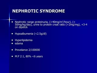
Nephrotic syndrome Nephrotic syndrome has historically been considered to include five principal clinical findings: 1. High-grade, albumin-dominant proteinuria (generally >3 to 3.5 g/day or spot urine protein/creatinine ratio of >3 to 3.5 [grams of protein per gram of creatinine]) 2. Hypoalbuminemia 3. Edema 4. Hyperlipidemia 5. Lipiduria Brenner
Nephrotic syndrome In practice, many clinicians refer to “nephroticrange” proteinuria regardless of whether their patients have the other manifestations of the full syndrome because the latter are consequences of the proteinuria.
Isolated proteinuria — Isolated proteinuria is defined as proteinuria without hematuria or a reduction in glomerular filtration rate (GFR). In most cases of isolated proteinuria, the patient is asymptomatic, and the presence of proteinuria is discovered incidentally by use of a dipstick during routine urinalysis. The urine sediment is unremarkable (fewer than three erythrocytes per high power field and no casts), protein excretion is less than 3 g/day (non-nephrotic), serologic markers of systemic disease are absent, and there is no hypertension, diabetes, and also no edema or hypoalbuminemia
Glomerularproteinuria — Glomerularproteinuria is due to increased filtration of macromolecules (such as albumin) across the glomerular capillary wall. This is a sensitive marker for the presence of glomerular disease. The proteinuria associated with diabetic nephropathy and other glomerular diseases, as well as more benign causes, such as orthostatic or exercise-induced proteinuria, fall into this category. Most patients with benign causes of isolated proteinuria excrete less than 1 to 2 g/day
Tubular proteinuria — Low-molecular-weight proteins, such as beta2-microglobulin, immunoglobulin light chains, retinol-binding protein, and polypeptides derived from the breakdown of albumin. These smaller proteins can be filtered across the glomerulus and are then almost completely reabsorbed in the proximal tubule. Interference with proximal tubular reabsorption, due to a variety of tubulointerstitial diseases or even some primary glomerular diseases, can lead to increased excretion of these smaller proteins .
Tubular proteinuria is often not diagnosed clinically since the dipstick for protein is not highly sensitive for the detection of proteins other than albumin and because the quantity of non-albumin proteins excreted is relatively low. The increased excretion of immunoglobulin light chains (or Bence Jones proteins) in tubular proteinuria is mild, polyclonal (both kappa and lambda), and not injurious to the kidney. This is in contrast to the monoclonal and potentially nephrotoxic nature of the light chains in the overflow proteinuria seen in multiple myeloma
Overflow proteinuria — Increased excretion of low-molecular-weight proteins can occur with marked overproduction of a particular protein, leading to increased glomerular filtration and excretion. This is almost always due to immunoglobulin light chains in multiple myeloma but may also be due to lysozyme (in acute myelomonocyticleukemia), myoglobin (in rhabdomyolysis), or free hemoglobin (in intravascular hemolysis) that is not bound to haptoglobin [6]. In these settings, the filtered load is increased to a level that exceeds the normal proximal reabsorptive capacity. Patients with myeloma kidney also may develop a component of tubular proteinuria since the excreted light chains may be toxic to the tubules, leading to diminished reabsorption.
Post-renal proteinuria — Inflammation in the urinary tract, which can occur with urinary tract infection, can give rise to increases in urinary protein excretion. The excreted proteins are generally non-albumin (often IgA or IgG), and only small amounts are excreted. Leukocyturia is frequently present in such patients. Patients with nephrolithiasis or tumors of the urinary tract may also have proteinuria.
NEPHROTIC SYNDROME • Pathophysiology • Proteinuria • Hypoalbuminemia • Edema • Hyperlipidemia • Cause (diagnosis and differential diagnosis) • Systemic renal disease hepatitis B associated glomerulonephritis, Henoch-Schonleinpurpura, systemic lupus erythematosus, diatetes mellitus, amyloidosis • Idiopathic nephrotic syndrome • Complications • Infection • Coagulation disorders • Protein malnutrition and dyslipidemia • Acute renal failure
Nephrotic syndrome (NS)results from increased permeability of Glomeulrar basement membrane (GBM) to plasma protein. It is clinical and laboratory syndrome characterized by massive proteinuria, which lead to hypoproteinemia ( hypo-albuminemia), hyperlipidemia and pitting edema.
Proteinuria • Proteinuria can be caused by systemic overproduction, tubular dysfunction, or glomerular dysfunction. It is important to identify patients in whom the proteinuria is a manifestation of substantial glomerular disease as opposed to those patients who have benign transient or postural (orthostatic) proteinuria.
Negative:<15mg/dL Trace: 15-30 mg/dL 1+: 30-100 mg/dL 2+: 100-300 mg/dL 3+: 300-1000 mg/dL 4+: >1000 mg/dL
Heavy proteinuria (albuminuria) Figure 3.
Hypoalbuminemia • Hypoalbuminemia is in part a consequences of urinary protein loss. It is also due to the catabolism of filtered albumin by the proximal tubule as well as to redistribution of albumin within the body. This in part accounts for the inexact relationship between urinary protein loss, the level of the serum albumin, and other secondary consequences of heavy albuminuria .
Under normal conditions, albumin production by the liver is12 to 14 g/day .Production equals the amount catabolized, predominantly in extrarenal locations. However, about 10% is catabolized in the proximal tubule of the kidney after reabsorption of filtered albumin. In patients with nephrotic syndrome hypoalbuminemia results from excessive urinary loss, decreased (inadequate)hepatic synthesis, and increased rates of albumin catabolism. Urinary albumin loss is an important contributor to the development of hypoalbuminemia. However, it is not a sufficient cause in most patients with nephrotic syndrome, becausethe rate of hepatic albumin synthesis can increase by at least threefold, thereby compensating for urinary albumin loss. Inadequate synthetic response to hypoalbuminemia by the liver. Enhanced loss of albumin in the gastrointestinal tract has also been proposed to contribute to hypoalbuminemia Brenner
Edema Edema is a major characteristic of nephrotic syndrome. The development of hypoalbuminemia reduces the oncoticpressure within the capillaries, and this favors the net translocation of fluid into the interstitial spaces. To the extent that this occurs, intravascular volume and blood pressure fall, and this triggers the sympathetic nervous system, activates the renin-angiotensin- aldosterone axis, elevates vasopressin levels, and modulates many other control systems that act together to promote net renal salt and water retention. This pathogenic sequence has been termed the underfill mechanism of salt and water retention in nephrotic syndrome.(Alb less than 1,GFR more than 75%)(M.C) Brenner
edema formation in many, perhaps most, nephrotic patients cannot be fully explained by underfill mechanisms. Although reduced intravascular oncotic pressures certainly exist in nephrotic patients, the net hydrostatic gradient for water movement across capillary beds is also influenced by the interstitial oncotic pressure, and this generally falls in parallel with reductions in plasma oncotic pressure. Consequently, the net hydrostatic pressure gradient from the intravascular compartment to the interstitial space may not significantly increase. Edema formation under these conditions may be the consequence of a primary form of renal salt and water retention. This pathogenic sequence for edema formation is called the overfill mechanism. (Alb more than 2,GFR less than %50) Brenner
Salt & water retention mechanisms: 1-increased Na/k/ATPase activity in CCT in basolateral membrane. 2-increased Na/H/exchanger activity . 3-ANP resistancydue to increased activity of PDE that lead to degredation of cGMP that is second messenger of ANP. 4-increased ENac activity
Hyperlipidemia • Most nephrotic patients have elevated levels of total and low-density lipoprotein (LDL) cholesterol with low or normal high-density lipoprotein (HDL) cholesterol . Lipoprotein (a) [Lp(a)] levels are elevated as well and return to normal with remission of the nephroticsyndrome.TG &VLDL & lipoproteins are increased. In lipid metabolism,decreased lipoprotein lipase(LPL)& decreased lecithin cholesterol acyltransferase(LCAT) & in lipid transport, increased apolipoprotein B100 & decreased hepatic uptake of LDLs. • Nephroticpatients often have a hypercoagulable state and are predisposed to deep vein thrombophlebitis, pulmonary emboli, and renal vein thrombosis. Brenner
Response to Hypoalbuminemia → reflex to liver --→synthesis of generalize protein ( including lipoprotein ) and lipid in the liver ,the lipoprotein high molecular weight can not loss in urine → hyperlipidemia Diminished catabolism of lipoprotein
Table 3a NEPHROTIC SYNDROME ASSOCIATED WITH SPECIFIC CAUSES (“SECONDARY” NEPHROTIC SYNDROME)
Table 3b NEPHROTIC SYNDROME ASSOCIATED WITH SPECIFIC CAUSES (“SECONDARY” NEPHROTIC SYNDROME)
Pathology patterns and clinical presentations of idiopathic nephrotic syndome
Renal biopsy • In adults, the nephrotic syndrome is a common condition leading to renal biopsy. In many studies, patients with heavy proteinuria and the nephrotic syndromes have been a group highly likely to benefit from renal biopsy in terms of a change in specific diagnosis, prognosis, and therapy. • Selected adult nephrotic patients such as the elderly have a slightly different spectrum of disease, but again the renal biopsy is the best guide to treatment and prognosis.
PRIMARY NEPHROTIC SYNDROME • Minimal Change Disease • Focal Segmental Glomerulosclerosis • Membranous Nephropathy • Membranoproliferative Glomerulonephritis (MPGN)
Minimal Change Nephropathy (MCN) The glomeruli appear normal basically Under Light microscopy, and Under Immunofluorescence. under Electron microscopy – fusion of the foot processes of the podocytes
Figure 5a. Pathology of glomerular disease. Light microscopy. (a) Normal glomerulus; minimal change disease.
PRIMARY NEPHROTIC SYNDROME • Minimal Change Disease • Focal Segmental Glomerulosclerosis • Membranous Nephropathy • Membranoproliferative Glomerulonephritis(MPGN)
Figure 5b. Segmental sclerosis; focal segmental glomerulosclerosis.
Pathology Light Microscopy: FSGS is characterized by focal and segmental glomerular sclerosis. The sclerosis may begin as segmental consolidation caused by insudation of plasma proteins causing hyalinosis, by accumulation of foam cells, by swelling of epithelial cells, and by collapse of capillaries resulting in obliteration of capillary lumens. These events are accompanied by an increase in extracellular matrix material that ultimately accounts for the sclerosis component of the lesion.
Immunofluorescence In all of the histologic variants, nonscleroticglomeruli and segments usually show nostaining for immunoglobulins or complement. A minority of patients with FSGS have low-level mesangial staining for IgM in nonscleroticglomeruli.
Electron Microscopy Nonscleroticglomeruli and segments should have no immune complex–type electron-dense deposits.