Download
1 / 24
320 likes | 695 Vues
Intro to the Liposome Project. liposomes. Advanced technology: Liposomes as drug delivery vesicles. O rganized structure allows liposomes to carry soluble or insoluble drugs. What are lipids ?

E N D
liposomes • Advanced technology: Liposomes as drug delivery vesicles. • Organized structure allows liposomes to carry soluble or insoluble drugs.
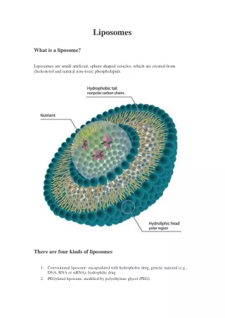
What are lipids? Lipidsare a group of chemical compounds (such as oils and waxes) which occur in living organisms and are only sparingly soluble in water
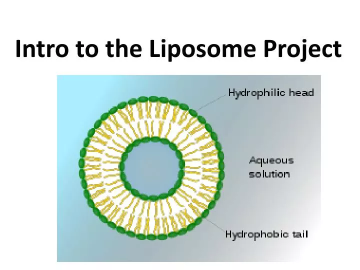
What are phospholipids? • Type of lipid that contains phosphate. • Phospholipids are the building blocks of liposomes and cell membranes. • Lipids in general are hydrophobic, also called non-polar (not able to be mixed in water). However, the phosphate group in phospholipids is hydrophilic, also called polar (able to be mixed in water).
Whenphospholipidsare immersed in water they arrange themselves so that their hydrophilic regions point toward the water and their hydrophobic regions point away from the water and stick together in bilayer form. The interaction betweenphospholipidsand water takes place at a temperature above the gel to liquid-crystalline phase transition temperature (TC) Which represents the melting point of the acyl chains.
When fully hydrated, most phospholipids exhibit a phase change from L-β gel crystalline to the L-α liquid crystalline state at TC. All phospholipids have a characteristic (TC), which depends on nature of the polar head group and on length and degree of unsaturation of the acyl chains. Above TC phospholipids are in the liquid-crystalline phase, characterized by an increased mobility the acyl chains. Decrease in temperature below (TC) induces transition to a more rigid state (Gel State) resulting in tightly packed acyl chains and the lipid molecules arrange themselves to form closed planes of polar head groups.
Liposomes can be formed from a variety of phospholipids. The lipid most widely used is phosphatidyl choline, phosphatidyl ethanolamime and phosphatidlyl serine either as such or in combination with other substance to vary liposome's physical, chemical and biological properties,liposome size, charge, drug loading capacity and permeability. Cholesterol: Condense the packing of phospholipids in bilayer above TC. Thereby reducing their permeability to encapsulated compounds. Stearylamine can be used to give positive charge to the liposomes surface.
Liposome Cell Membrane Phospholipid Bilayersare the core structure ofliposomeand cell membrane formations. Thus the structure ofliposomesis similar to the structure of cell membranes.
Liposomescan contain and mobilize water-soluble materials as well as oil-soluble materials in specific cavities inside themselves .
Micropipette P-1000 P-200
Waste today… Pipette tips can go in garbage. Put glass slides in glass waste. Extra liposome solution? Put in waste container in fume hood.
Synthesis of Liposomes Procedure: • Obtain lipid/chloroform solution from Avanti lipids. • Add 20 drops (using a glass pipette) to a glass vial. • Make a solution of fat soluble dye (such as oil red) in chloroform or another non-polar solvent. It should have a concentration of 0.2 mg/mL. Add 500 µL of the dye solution to your glass vial with the lipids. • Let the chloroform/oil red/lipids solution dry out overnight so the lipid & dye are close together. • Next, form liposomes by adding 400 µL water. Vortex or sonicate. • Spin down solution with a mini-centrifuge—the liposomes stay at the top and excess dye/junk goes to the bottom. • Pipette the liposomes onto a glass slide and place another glass slide on top of the solution. • Use ExoLab cameras, microscopes & iPads to measure the diameter of the liposomes. • You may also choose to use a mini-extruder with various filters to make your liposomes smaller. I already did the part in red, only you are not working with lipids + dye today, just lipids.
Next, form liposomes by adding 400 µL distilled water. Vortex or sonicate. • Start by vortexing for 30 seconds, then wait 5 minutes, then vortex again for another 30 seconds, then wait 5 minutes. 6. Spin down solution with a mini-centrifuge—the liposomes stay at the top and excess dye/junk goes to the bottom. • 7. Pipette 30 µL of the liposomes onto a glass slide and place another glass slide a coverslip on top of the solution. • 8. Use ExoLab cameras, microscopes & iPads to measure the diameter of the liposomes. • 9. You may also choose to use a mini-extruder with various filters to make your liposomes smaller.