Download
1 / 69
690 likes | 795 Vues
Bridget, Jephte, Kristi, Matt, Teresa Set up 20 µl mix for each primer/DNA combo on ice! 2 µl 10x F primer (1 pMol/µl = 1µM final []) 2 µl 10x R primer 1 µl DNA We will prepare Phusion master mix for 130 µl total volume 26 µl 5x Phusion HF buffer 2.6 µl 10 mM dNTP (200 µM final [])

E N D
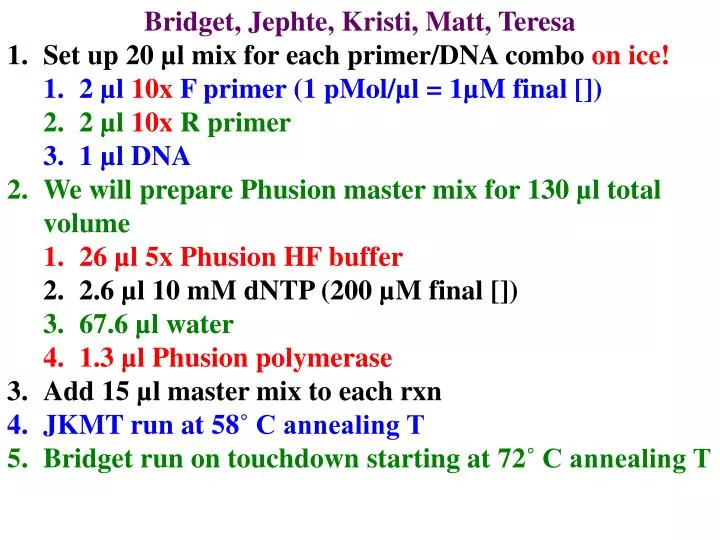
Bridget, Jephte, Kristi, Matt, Teresa • Set up 20 µl mix for each primer/DNA combo on ice! • 2 µl 10x F primer (1 pMol/µl = 1µM final []) • 2 µl 10x R primer • 1 µl DNA • We will prepare Phusion master mix for 130 µl total volume • 26 µl 5x Phusion HF buffer • 2.6 µl 10 mM dNTP (200 µM final []) • 67.6 µl water • 1.3 µl Phusion polymerase • Add 15 µl master mix to each rxn • JKMT run at 58˚ C annealing T • Bridget run on touchdown starting at 72˚ C annealing T
Testing internal primers • Set up 20 µl mix for each primer/DNA combo on ice! • 2 µl 10x internal F primer (1 pMol/µl = 1µM final []) • 2 µl 10x internal R primer • 0.5 µl DNA = best PCR reaction • We will prepare master mix for 90 µl total volume • 9 µl 10x Taq buffer • 1.8 µl 10 mM dNTP (200 µM final []) • 58.4 µl water • 0.56 µl Taq polymerase • Add 15.5 µl master mix to each rxn • Run at 50˚ C annealing T, 2 minutes/72˚ cycle
2 Protein Targeting pathways Protein synthesis always begins on free ribosomes In cytoplasm 1) Post -translational: proteins ofplastids,mitochondria, peroxisomesandnuclei 2) Endomembrane system proteins are imported co-translationally
Sorting proteins made on RER lysosomal proteins are targeted by mannose 6-phosphate M 6-P receptors in trans-Golgi direct protein to lysosomes (via endosomes) M 6-P is added in Golgi by enzyme that recognizes lysosomal motif
Glycosylation within ER All endomembrane proteins are highly glycosylatedon lumenal domains. Glycosylation starts in the ER, continues in the Golgi
Glycosylation within ER • All endomembrane proteins are highly glycosylatedon lumenal domains. • Glycosylation starts in ER, continues in Golgi • makes proteins more hydrophilic • essential for proper function • tunicamycin poisons cells • Glycosylation mutants are even sicker
Glycosylation in RER 1)(CH2O)n are assembled stepwise on dolichol-PO4 2) Transfer (CH2O)n to target asn
Glycosylation in RER 1)(CH2O)n are assembled stepwise on dolichol-PO4 2) Transfer (CH2O)n to target asn 3) remove 2 glucose & bind chaperone If good,remove gluc 3 & send to Golgi
Glycosylation in RER remove 2 glucose & bind to chaperone If good,remove gluc 3 & send to Golgi If bad, GT adds glucose & try again Eventually, send bad proteins to cytosol & eat them
Glycosylation • next modify (CH2O) n in Golgi • Remove some sugars & add others
Glycosylation • next modify (CH2O) n in Golgi • Remove some sugars & add others • different rxns occur in different parts of Golgi • why we separate Golgi into distinct regions
Post-translational protein targeting Key features 1) imported after synthesis
Post-translational protein targeting Key features 1) imported after synthesis 2) targeting information is motifs in protein a) which organelle b) site in organelle 3) Receptors guide it to correct site 4) no vesicles!
Protein targeting in Post-translational pathway • SKL (ser/lys/leu) at C terminustargets most peroxisomal matrix proteins = PTS1 • In humans 3 are targeted by 9 aa at N terminus = PTS2 • Defective PTS2 receptor causes Rhizomelic chondrodysplasia punctata N SKL C N PTS2 C
Targeting peroxisomal proteins • Bind receptor in cytoplasm • Dock with peroxisomal receptors • Import • protein w/o • unfolding it! • Recycle • receptors
Peroxisomal Membrane Synthesis Most peroxisomes arise by fission can arise de novo! Mechanism is poorly understood/ may involve ER! Only need PEX 3 & PEX 16 to import pex membrane prot
Protein import into nuclei • nuclear proteins are targeted by internal motifs • necessary & sufficient to target cytoplasmic proteins to nucleus
Protein import into nuclei • nuclear proteins are targeted by internal motifs • as in golgi, are not specific • shapes cf sequences • Receptors bind objects of the right shape!
Protein import into nuclei • 3 types of NLS (nuclear localization sequence) • 1) basic residues in DNA-binding region + + + LZ
Protein import into nuclei • 3 types of NLS (nuclear localization sequence) • 1) basic residues in DNA-binding region • 2) SV-40 KKKRK + + + LZ KKKRK
Protein import into nuclei • 3 types of NLS (nuclear localization sequence) • 1) basic residues in DNA-binding region • 2) SV-40 KKKRK • 3) bi-partite: 2-4 basic aa,10-20 aa spacer, 2-4 basic aa + + + LZ KKKRK + + + +
Protein import into nuclei • 1) importin-a binds NLS importin-b binds complex • 2) escort to nuclear pores • Pores decide who can enter/exit nucleus
Protein import into nuclei 1) importin-a binds NLS, importin-b binds complex 2) escort to nuclear pores 3) transporter changes shape, lets complex enter 4) nuclear Ran-GTP dissociates complex 5) Ran-GTP returns b-importinto cytoplasm, becomes Ran-GDP. GTP -> GDP = nuclear importenergy source 6) Exportins return a-importin& other cytoplasmic prot
Protein import into cp and mito • Many common features • 1) Pulse-chase experiments show most cp & mt proteins are made in cytoplasm as larger precursor (preprotein) • both have N-terminal targeting peptide • transit peptidein cp • presequence in mito • necessary & sufficient to target
Protein import into cp &mito • Many common features • 1) N-terminal transit peptideor presequence • necessary & sufficient to target • usually removed upon arrival
Protein import into cp & mito • Many common features • 1) N-terminal transit peptideor presequence • 2) both need energy input • a) ATP for both • b) Mt also use Proton Motive Force (PMF) • H+ gradient made by electron transport • c) Cp also use GTP (but not PMF)
Protein import into cp & mito • 1) N-terminal transit peptideor presequence • 2) both need energy input • 3) proteins unfold to enter, then refold inside • a) need chaperonins on both sides of membrane • i) chaperonins in cytosol unfold • ii) chaperonins inside refold • a) helps draw through membrane
Protein import into mitochondria • Precursor has N-terminal targeting presequence • 20 - 70 aa • 1. Many basic a.a (+ charge) = lys, arg • 2. Many hydroxylated a.a. (ser, thr) • 3. Segment can fold into a-helix + + + presequence mature protein presequence
Protein import into mitochondria 1) HSP70 binds & unfolds preprotein
Protein import into mitochondria 1) HSP70 binds & unfolds preprotein 2) Unfolded presequence binds MOM receptors (MOM19 & MOM72)
Protein import into mitochondria 1) HSP70 binds & unfolds preprotein 2) Unfolded presequence binds MOM receptors 3) Unfolded protein is translocated through MOM controversy: do inner and outer membrane contact each other before protein import?
Protein import into mitochondria 1) HSP70 binds & unfolds preprotein 2) Unfolded presequence binds MOM receptors 3) Unfolded protein is translocated through MOM 4) Unfolded protein is translocated through MIM presequence contacts MIM proteins
Protein import into mitochondria 5) Chaperones in matrix refold protein 2 different chaperones: mHSP70 & HSP60 consumes ATP
Protein import into mitochondria Driving forces for import: 1) PMF (on +ve a.a.) 2) Refolding (Brownian ratchet) 3) ATP hydrolysis used to drive unfolding and refolding
Protein import into mitochondria 6) Once protein is refolded, targeting sequence is removed
CP protein import • chaperones in cytoplasm unfold preprotein • transit peptide contacts receptor in OE • transit peptides:longer & less +ve than presequences • Just a few changes convert transit peptide to presequence
CP protein import • 1) chaperones in cytoplasm unfold preprotein • 2) transit peptide contacts receptor in OE • 3) Unfolded protein is translocated through OE • requires GTP • difference from mito
CP protein import 1) chaperones in cytoplasm unfold preprotein 2) transit peptide contacts receptor in OE 3) Unfolded protein is translocated through OE 4) Unfolded protein is translocated through IE
CP protein import 5) Protein is folded on inside by chaperones 6) transit peptide is removed
Energy for cp import 1) GTP hydolysis:crossing OE 2) Refolding (Brownian ratchet) 3) ATP hydrolysis: un- & refolding
CP Protein import Targeting to other parts requires another motif Hypothesis: proteins enter stroma first, then find their final destination
Proteomics • studying all of the proteins present in a particular organism • Now that we have the genome, what do we do with it? • old way was to prepare 2-D • gels of proteins prepared from • the cells being studied • first use isoelectric focusing • to separate proteins by pI
Proteomics • old way was to prepare 2-D gels of proteins prepared from the organisms or tissues being studied • first use isoelectric focusing to separate proteins by pI • Then use SDS-PAGE to separate by size
Proteomics • old way was to prepare 2-D gels of proteins prepared from the organisms or tissues being studied • first use isoelectric focusing to separate proteins by pI • Then use SDS-PAGE to separate by size
Proteomics • Use 2D-SDS-PAGE to • Separate by pI then size • Then ID each spot! • Sequencing
Proteomics • Use 2D-SDS-PAGE to • Separate by pI then size • Then ID each spot! • Sequencing • Slow (1aa/hr)
Proteomics • Use 2D-SDS-PAGE to • Separate by pI then size • Then ID each spot! • Sequencing • Slow (1aa/hr) • 98% accurate = 50 aa • limit
MALDI (matrix assisted laser desorption ionization) to ionize peptides so they can be analyzed by mass spectrometry. • sample is dispersed in a large excess of matrix material which absorbs the incident laser
sample is dispersed in a large excess of matrix material which absorbs the incident laser • Short pulses of laser light focused on the sample cause the sample and matrix to volatilize