Download
1 / 4
40 likes | 129 Vues
F ig. S1 Characterization of RLs. (A). 3. 5. 6. 1. 2. 4. (B). E CM. 2 & 4. 3. 6. 1. 5.

E N D
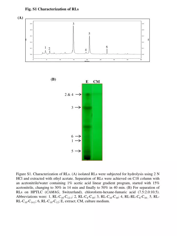
Fig. S1 Characterization of RLs (A) 3 5 6 1 2 4 (B) E CM 2 & 4 3 6 1 5 Figure S1. Characterization of RLs. (A) isolated RLs were subjected for hydrolysis using 2 N HCl and extracted with ethyl acetate. Separation of RLs were achieved on C18 column with an acetonitrile/water containing 1% acetic acid linear gradient program, started with 15% acetonitrile, changing to 30% in 14 min and finally to 50% in 40 min. (B) For separation of RLs on HPTLC (CAMAG, Switzerland), chloroform-hexane-fumaric acid (7.5:2.0:10.5). Abbreviations were: 1, RL-C10-C12:1; 2, RL-C8-C10; 3, RL-C10-C10; 4, RL-RL-C8-C10 ; 5, RL-RL-C10-C14:1; 6, RL-C10-C12; E, extract; CM, culture medium.
Fig. S2 Stability of nanoparticles $ % & Figure S2. Stability assessment of bare ZnO and RL@ZnO nanoparticles up to 15 months of the storage. Aqueous solutions (200 µg/mL) of both bare ZnO and RL@ZnO nanoparticles were prepared and subjected for the change in UV–vis spectrum at A360 nm. Bar graph shows the month-wise absorbance of both types of nanoparticles. Data represent mean SD. Different letters: $, %, and & significantly different from their respective control according to post hoc comparison (LSD-test) (P < 0.01).
Fig. S3 Antioxidant activity of bare ZnO nanoparticles (B) (A) (C) (D) (E) Figure S3. DPPH radical scavenging activity of bare ZnOnanoparticles. Various concentrations (0, 25, 50, 100, 150, & 200 mg/mL) of bare ZnO nanoparticles were added to freshly prepare stable DPPH solution (6 x 10⁻⁵M), mixed and incubated at room temperature for 60 min. Then, DPPH radical scavenging potential was monitored by UV-visspectrophotometer in the range of A460600 nm wavelength. DPPH radical scavenging activity was measured at (A) 3, (B) 6, (C) 9, (D) 12 and (E) 15 months of storage.
Fig. S4 Antioxidant activity of nanoparticles (A) @ # $ % @ & # $ @ @ # $ # % (B) @ $ # # % @ & $ % @ # $ @ # % $ & Figure S4. Inhibition of lipid peroxidation (LPO) and O2• radical anion free radical scavenging by RL@ZnO nanoparticles. (A) Aqueous egg yolk homogenate (10%), indicated concentrations of nanoparticles and 0.07 M FeSO4 were added and incubated at 37 0C for 30 min to induce lipid peroxidation. Thereafter, 20% acetic acid, 0.8% TBA in 1.1% sodium dodecyl sulphate and 20% TCA were mixed added, heated in a boiling water bath for 30 min. After cooling, reaction mixture was extracted with butan-1-ol and recorded the absorbance at A532 nm. (B) The riboflavin-light-NBT system was used to determine the O2• radical scavenging potential. The mixture contained (50 mM phosphate buffer; pH 7.8; 13 mM methionine, 2 µM riboflavin, 100 µM EDTA, 75 µM NBT) and indicated concentrations of bare ZnO and RL@ZnO nanoparticles. It was kept in front of fluorescent light for 10 min and absorbance was measured at A560 nm. Data represent mean SD. Different letters: @, #, $, %, and & significantly different from their respective control according to post hoc comparison (LSD-test) (P < 0.01).