Download
1 / 14
150 likes | 428 Vues
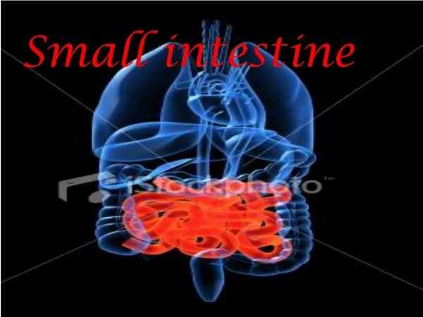
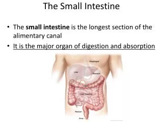
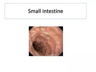
Small Intestine. The small intestine, is a convoluted tube, about 6 meters long, extending from the pylorus to the ileo-cecal valve , situated centrally in the abdominal cavity and is flanked laterally and superiorly by the large intestine.

E N D
The small intestine, is a convoluted tube, about 6 meters long, extending from the pylorus to the ileo-cecal valve, situated centrally in the abdominal cavity and is flanked laterally and superiorly by the large intestine.
Divisions:1. Duodenum 2. Jejunum 3. Ileum • Jejunum and ileum They form the mobile part of the small intestine, suspended from the posterior abdominal wall by the mesentery.
Jejunum Ileum • Length: proximal 2/5 (8 feet) distal 3/5 (12 feet) • Diameter: Wide Narrow • Arterial arcades: Few and simple Numerous (3-4), complex
Jejunum Ileum 4- Mesentery: Few fat,+ve windows Much fat, no windows 5. Mucosal Circular Folds: Numerous Few ( Plica Circularis)
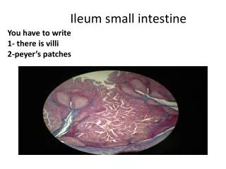
Jejunum Ileum 6- Wall : thick thin 7-Pyer’s patches: -ve +ve
Mesentery of the small intestine: is a fan- shaped peritoneal fold which has an anterior free border and posterior attached border. The anterior border contains the jejunum and ileum and is 6 meter long. The posterior border is the root of mesentery and is 6 inches long.
Structures crossed by the root of the mesentery (6): • 3rd part of duodenum • Abdominal aorta and right gonadal vessels. • IVC. Right psoas major. Right ureter. • Right genito-femoral nerve.
Contents of the mesentery: • Superior mesenteric artery. • Superior mesenteric vein. • Coils of the small intestine • Extraperitoneal tissue and fat. • Sympathetic nerve fibers. • Mesenteric LN (arranged in three groups: large, medium, and small).
Superior mesenteric artery • Origin: from descending abdominal aorta 1cm below coeliac trunk at the level of lower border of L1 vertebra
Course: • It is accompanied by superior mesenteric vein on its right side and both of them run in the root of mesentery.
Structures supplied by SMA: It supplies midgut .
Branches of sup. mesenteric a. • inferior pancreaticoduodenal artery Supplieshead of the pancreas and to the descending and inferior parts of the duodenum • middle colic artery Supplies to the transverse colon • right colic artery to ascending colon • intestinal arteries branches to ileum, jejunum • ileocolic artery (terminal branch of the SMA) supplies last part of ileum, cecum, and appendix
Thank You Prof.: Dr. Shawky Tayel