Download
1 / 37
370 likes | 373 Vues
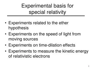
This chapter explores the discovery of X-rays and electrons, the phenomenon of line spectra, and the experimental basis for the quantum theory. Topics include the photoelectric effect, blackbody radiation, X-ray production, and more.

E N D
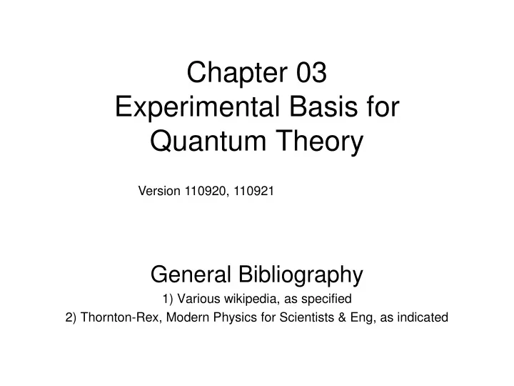
Chapter 03Experimental Basis for Quantum Theory General Bibliography 1) Various wikipedia, as specified 2) Thornton-Rex, Modern Physics for Scientists & Eng, as indicated Version 110920, 110921
Outline • 3.1 X-rays and Electrons • 3.2 Electron Charge • 3.3 Line Spectra • 3.4 Quantization • 3.5 Blackbody Radiation • 3.6 Photoelectric Effect • 3.7 X-Ray Production • 3.8 Compton Effect • 3.9 Pair Production & Annihilation
3.1 X-rays and Electrons http://physics.kenyon.edu/EarlyApparatus/Static_Electricity/Geissler_Tubes/Geissler_Tubes.html http://www.oneillselectronicmuseum.com/page9.html http://www.beer-neon-signs.com/category/neon-business-signs Geissler tubes ~1857
3.1 X-rays and Electrons Discovered cathode rays could be shifted by a magnet http://www.magnet.fsu.edu/education/tutorials/museum/crookes_tube.html Cathode rays (now called electrons) Crookes Tubes ~1869
X-rays and Electrons Hand mit Ringen (Hand with Rings): print of Wilhelm Röntgen's first "medical" X-ray, of his wife's hand, taken on 22 December 1895 and presented to Ludwig Zehnder of the Physik Institut, University of Freiburg, on 1 January 1896[17][18]
X-rays and Electrons 1896 plaque published in "Nouvelle Iconographie de Salpetriere", a medical journal. In the left a hand deformity, in the right same hand seen using radiography. The authors designated the technique as Röntgen photography.
X-rays and Electrons JJ Thomson http://www.scifun.ed.ac.uk/pages/pp4ss/pp4ss-eoverm.html http://ixnovi.people.wm.edu/Onebeautifulexperiment2008/emexperimentbywilliamsendor3.html
3.3 Line Spectra http://nothingnerdy.wikispaces.com/DIFFRACTION+GRATING Pink Floyd: Dark Side of the Moon http://intro.chem.okstate.edu/1314f00/Lecture/Chapter7/Lec11300.html
k = 3,4,5,6 http://www.chem1.com/acad/webtext/atoms/atpt-3.html
3.3 Line Spectra Lyman n=1 Brackett n=4 Paschen n=3 Pfund n=5 Balmer n=2 1000 nm 100 nm vis 10000 nm
3.5 Blackbody Radiation http://www.mytightride.com/fof1fefl.html
3.5 Blackbody Radiation http://en.wikibooks.org/wiki/Wikijunior:How_Things_Work/Light_Bulb http://www.freefoto.com/preview/11-12-52/Electric-Light-Bulb http://phet.colorado.edu/sims/blackbody-spectrum/blackbody-spectrum_en.html
3.5 Blackbody Radiation Wein’s Law Stefan-Boltzmann Law e = 1 for perfect blackbody
3.5 Blackbody Radiation UV IR
3.5 Blackbody Radiation Max Planck assumed some sort of oscillators filled the cavity AND energy difference between standing wave modes = h f Planck’s Radiation Law h = 6.626e-34 Js
3.6 Photoelectric Effect http://en.wikipedia.org/wiki/Heinrich_Hertz http://en.wikipedia.org/wiki/Induction_coil Heinrich Hertz Verified Maxwell equations prediction of electromagnetic waves ~1887
3.6 Photoelectric Effect Photoelectric Effect Investigations ~1900 1. Higher intensity light did not change the point at which current started to flow. (i.e. energy of the electrons) 1’. More total incident energy did not increase the energy of individual electrons. 2. Different colors of light changed the starting point for current flow.
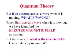
3.6 Photoelectric Effect Energy of an individual photon Work function typically a few Volts at most Implications: 1. EM waves have fixed energies (EM field is quantized) 2. Electrons are bound in a material by an amount determined by the composition
3.7 X-Ray Production(Bremsstralung + Characteristic X-Rays) At fixed HV 35kV Roentgen characteristic lines min l max Energy
3.7 X-Ray Production(Bremsstralung + Characteristic X-Rays) Roentgen
3.7 X-Ray Production(Bremsstralung + Characteristic X-Rays) Do TV Sets Give Off X-Rays? X-rays may be produced when electrons, accelerated by high voltage, strike an obstacle while traveling in a vacuum, as in a TV containing a cathode ray tube (CRT). Since many of the components in television sets operate at thousands of volts, there is the potential for x-ray generation. These components may produce x-rays capable of escaping from the television receiver or CRT. This unintentional emission of x-radiation can pose a potential hazard and must be controlled. http://en.wikipedia.org/wiki/Cathode_ray_tube http://www.fda.gov/Radiation-EmittingProducts/ResourcesforYouRadiationEmittingProducts/ucm252764.htm
http://www.orau.org/ptp/collection/xraytubescoolidge/xraytubescoolidge.htmhttp://www.orau.org/ptp/collection/xraytubescoolidge/xraytubescoolidge.htm
http://www.tradevv.com/chinasuppliers/eiffelgu_p_23e13/china-Portable-X-ray-flaw-detector-ceramic-tube.htmlhttp://www.tradevv.com/chinasuppliers/eiffelgu_p_23e13/china-Portable-X-ray-flaw-detector-ceramic-tube.html http://www.aerospacendt.com/Radiography.htm
Computed tomography (CT) scanning, also called computerized axial tomography (CAT) scanning, is a medical imaging procedure that uses x-rays to show cross-sectional images of the body. A CT imaging system produces cross-sectional images or "slices" of areas of the body, like the slices in a loaf of bread. These cross-sectional images are used for a variety of diagnostic and therapeutic purposes. How a CT system works: http://www.strokecenter.org/patients/diagnosis/ct.htm http://www.fda.gov/Radiation-EmittingProducts/RadiationEmittingProductsandProcedures/MedicalImaging/MedicalX-Rays/ucm115317.htm
http://medicaltools.onsugar.com/Ct-Scan-Abdomen-Cancer-15819123http://medicaltools.onsugar.com/Ct-Scan-Abdomen-Cancer-15819123 http://www.strokecenter.org/patients/diagnosis/ct.htm http://info.shields.com/bid/43193/MRI-Images-torn-ACL-and-normal-ACL
Fluoroscopy is an imaging technique commonly used by physicians to obtain real-time moving images of the internal structures of a patient through the use of a fluoroscope. In its simplest form, a fluoroscope consists of an X-ray source and fluorescent screen between which a patient is placed. However, modern fluoroscopes couple the screen to an X-ray image intensifier and CCDvideo camera allowing the images to be recorded and played on a monitor. http://en.wikipedia.org/wiki/Fluoroscopy
3.8 Compton Effect Thomson Scattering In classical description, scattering occurs via dipole and scattered photon of same frequency (wavelength)
3.8 Compton Scattering h/mc =2.2426e-12 m
3.9 Pair Production & Positron Annihilation Pair Production
3.9 Pair Production & Positron Annihilation Positron Annihilation Photons come out back-to-back Photon energies are 0.511 MeV each
Positron emission tomography (PET) is a nuclear medicineimaging technique that produces a three-dimensional image or picture of functional processes in the body. The system detects pairs of gamma rays emitted indirectly by a positron-emitting radionuclide (tracer), which is introduced into the body on a biologically active molecule. Three-dimensional images of tracer concentration within the body are then constructed by computer analysis. If the biologically active molecule chosen for PET is FDG, an analogue of glucose, the concentrations of tracer imaged then give tissue metabolic activity, in terms of regional glucose uptake. http://en.wikipedia.org/wiki/PET_scanner
Summary of Chapter 03Strange things not known from classical physics • New things • Cathode rays electrons • X-rays • Line spectra • Gaseous discharges show lines rather than continous spectrum • Blackbody radiation • Rayleigh-Jeans classical formula clearly incorrect at explaining spectrum • Planck: oscillators with fixed energies • Compton scattering • Scattered photons have different wavelength in contast to classical description • Pair production & Positron annihilation • Waves change into particles and vice versa