Download
1 / 49
560 likes | 1.68k Vues
Syncope in Elderly SYNCOPE Syncope (Greek: synkope = cut-off) is a brief transient loss of consciousness (fainting) and postural tone (collapse) with rapid spontaneous recovery SYNCOPE From 1997-2000 National Health Ambulatory Medical Survey of ED visits in USA.

E N D
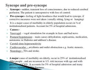
SYNCOPE • Syncope (Greek: synkope = cut-off) is a brief transient loss of consciousness (fainting) and postural tone (collapse) with rapid spontaneous recovery
SYNCOPE • From 1997-2000 National Health Ambulatory Medical Survey of ED visits in USA. • 2.63 million ED pt.( 0.65% of all visits) with the diagnosis of syncope unrelated to injury • 1.1 million pt.(40.8%) were 65 yrs or older • 63.8% were female • Among pt. older than 65 yrs, admit rate for syncope was 61.8% and was the sixth most common admission diagnosis
SYNCOPE • Age-dependent morphological and physiological changes • Old patients often take drugs (sedatives, diuretics, vasodilators, anti-hypertensives) • Old patients display a higher incidence of chronic pathologies such as diabetes mellitus, congestive heart failure, coronary disease, cerebrovascular pathologies and multiple sensorial deficiency.
Age-related physiological changes that predispose to syncope • Blood vessel • Heart • Autonomic nervous system • Other non cardiological changes
Blood vessel • Atherosclerosis is also universally present in older humans • Impair endothelial-dependent nitric oxide release • Increase endothelin release in the ageing vessels • This impairs both the cardiac and cerebral circulation which may predispose to syncope in the elderly.
Heart • Age-related stiffening of arterial vessels produces high afterload. • Ventricular walls become more fibrotic and noncompliant leading to ventricular diastolic dysfunction. • LV systolic dysfunction is also common because of the high prevalence of HTN and IHD among the elderly • Increase incidence of age-related mitral and aortic valvular diseases.
Heart • A progressive fall in the ratio of nodal myocytes to collagenous stroma with age particularly in the SA node increases the incidence of AF, heart block and sick sinus syndrome.
Autonomic nervous system • Beta-adrenergic response to plasma noradrenaline is blunted in the elderly • Diminished beta-1 responses lead to reduced cardioacceleration and cardiac contractility • Diminished beta-2 results in increased vascular tone because of the unopposed alpha-1 vasoconstriction. • Baroreflex mediated cardioacceleration is also reduced
Autonomic nervous system • HR increase in response to stress is less effective. • Sympathetic and parasympathetic autonomic responses are reduced in health ageing • Blunted autonomic responses together with other factors including dehydration, vasodilator medications, sodium wasting may result in orthostatic hypotension, cerebral underperfusion and syncope in the elderly.
Other non cardiological change • Plasma renin and aldosterone fall with age and this results in sodium wasting. • impaired thirst response of many elderly people to hyperosmolality may cause hypovolaemia and consequent orthostatic hypotension
Causes of syncope of the elderly • Cardiac diseases - Primary cardiac arrhythmias - Structural cardiovascular diseases—obstruction to left ventricular outflow - Obstruction to right ventricular outflow • Neurally mediated syncopal syndromes - Vasovagal syncope - Situational syncope - Carotid sinus hypersensitivity • Orthostatic and dysautonomic disturbance of blood pressure control • Postprandial hypotension • Cerebrovascular, neurological, and psychiatric causes
Primary cardiac arrhythmias • Probably the most common cause of syncope in patients with structural heart or vascular disease. • An age-related fall in nodal myocytes particularly in the sino-atrial node increases the incidence of atrial fibrillation, heart block and sick sinus syndrome • Polypharmacy
Drugs predisposing to syncope • Vasodilators : nitrates, CCB, hydralazine, ACEIs • AntiHT : clonidine, BB • Prolongation of QT(torsade de pointes) - Antiarrhythmic agent : class IA,III - ATB : macrolide(erythromycin), bactrim - Others : terfenadine,TCA, cisapride, phenothiazines, probucol
Conditions predisposing to a prolonged QT interval and torsade des pointes
Structural cardiovascular diseases obstruction to left ventricular outflow • Aortic stenosis is the most common structural lesion associated with syncope in the elderly - Age < 70 yr.: Congenital bicuspid valves - Age > 70 yr.: Degenerative changes • Hypertrophic obstructive cardiomyopathy (HOCM) • Vasodilator drugs or even vasodilatation after a hot bath can induce syncope in these patients
Obstruction to right ventricular outflow • The limitations to right ventricular outflow may lead to diminished capacity to increase cardiac output. • 18% of elderly pts admitted to an acute geriatric ward had pulmonary embolism in one study (Impallomehi et al., 1995) • Myoxma, pulmonary stenosis and pulmonary hypertension
Vasovagal syncope The mechanism of tilt or haemorrhage-induced vasovagal syncope
Situational syncope • Peripheral receptors similar to ventricular mechanoreceptors are found in lung, bladder, GI tract • Cough or micturition related syncope
Carotid sinus hypersensitivity • 20% of older people who presented with unexplained syncope (Parry and Eltrafi). • Defined as asystole of 3 s or more and/or a decrease in systolic pressure of 50 mmHg or more during carotid sinus massage.
Orthostatic and dysautonomic disturbance of BP control • 30% of community-dwelling adults over 75 years of age have orthostatic hypotension (Lipsitz, 1989). • Autonomic failure such as multiple system atrophy and diabetes mellitus. • The combination of the blunted age-related autoregulatory changes, medications (diuretic, vasodilators), and chronic diseases predispose older adults to orthostatic hypotension.
Postprandial hypotension • 8% of syncope cases in older nursing home patients in one study (Jansen et al., 1995). • Defined as 20 mmHg or greater decline in systolic blood pressure within 90 min after a meal. • Common in older adults and can coexist with orthostatic hypotension in the same individual ( Jansen and Lewis, 1995). • Pathophysiological mechanism of postprandial hypotension is still a matter of debate.
Cerebrovascular, neurological, and psychiatric causes • Syncope is rarely due to cerebrovascular disease unless there are accompanying focal neurological deficits. • Transient posterior circulation ischaemia can result in loss of consciousness and there are usually brain stem signs present including diplopia, vertigo, dysarthria, or hemiparesis. • Vasovagal syncope may mimic seizures • Psychiatric disturbances including hysterical reaction, panic attack with hyperventilation can either mimick or may lead to true syncope
Diagnostic evaluation • An emergency physician, when faced with a syncope-patient in an ED setting, should first seek to exclude life-threatening causes of syncope, which require immediate diagnostic evaluation/treatment + hospital admission
Diagnostic evaluation • AMI • PE • aortic dissection • cardiac tamponade • tension pneumothorax • leaking AAA • active internal bleeding • malignant cardiac arrhythmias • SAH • carotid artery/vertebral artery dissection
Diagnostic evaluation • If there are no overt life-threatening causes of syncope, then an emergency physician should attempt to identify patients with situational syncope, vasovagal syncope and benign orthostatic (postural) syncope - who are candidates for home discharge after any necessary stabilization treatment in the ED • If the cause of the syncope is not readily apparent after initial clinical evaluation in the ED, then an emergency physician should attempt to decide whether certain categories of syncope-patients require admission to hospital
History • An eye-witness account is very important • mode of onset and progression of event • Body position at onset of event • Depth of altered consciousness • Duration of the syncopal episode • Rate of recovery of consciousness • Identify any precipitants including meals, pain, cough, micturition, defaecation, swallowing, postural change, neck movement and exercise
History • Associated symptoms such as palpitation, dyspnoea, chest pain • History of panic attack and hyperventilation • pscyhological triggering events (painful stimuli, sudden bad news) • Drug history is obviously important. • Past medical history and risk factors for ischaemic heart disease
Physical examination • Search for trauma and assessment of severity • Cardiovascular examination - BP - Pulse volume - Neck bruits - JVP - Apex beat, Heart sounds • Abdomen • Neuro exam
BP • Difference in BP between lt. and rt. upper limbs > 20mmHg is abnormal (suggests dissecting aortic aneurysm or subclavian steal syndrome) • Difference in BP between upper and lower limbs > 20mmHg when recumbent is abnormal (suggests a dissecting aortic aneurysm) • Orthostatic vital signs : positive test is defined as a SBP decrease of > 20 - 30mmHg, a DBP decrease of >10 - 15mmHg and/or HR increase of greater than 30 bpm when standing
BP • A significant drop in BP + fixed HR suggests dysautonomia • A significant drop in BP + increased HR suggests volume depletion and/or excessive vasodilatation • An insignificant drop in BP + marked increase in HR suggests postural tachycardia syndrome(history of frequent fainting, symptoms of autonomic overactivity - palpitations, diaphoresis, tremulousness, visual blurring, non-anginal chest pain, "spaced-out" feelings, inability to concentrate, inability to breathe, sensations of impending doom)
Pulse volume • Decreased and delayed upstoke (aortic stenosis/hypertrophic obstructive cardiomyopathy) • Positive pulsus paradoxus (cardiac tamponade, massive pulmonary embolism) • Absent pulses (dissection of the aorta, cardiac emboli)
Heart sounds • Decreased (pericardial tamponade) • 3rd/4th heart sounds (ventricular failure or LV overload) • Loud second heart sound (pulmonary embolism or pulmonary hypertension) • Ejection systolic murmurs (aortic stenosis or hypertrophic cardiomyopathy - increased murmur when standing, decreased when squatting) • Machinary murmur (air embolism) • "tumor plop" or diastolic murmur (atrial myxoma) • Varying heart sounds/murmurs (thrombotic occlusion of a prosthetic valve)
Abdomen • Pulsatile masses (abdominal aneurysm) • Rectal exam for melena or heme-occult positive stools (gastro-intestinal bleeding) • Absent/decreased femoral pulses (dissection of the aorta)
Investigation • EKG • Echocardiography • Carotid sinus massage • Exercise stress test • Ambulatory continuous electrocardiography • Event and memory loop ambulatory electrocardigram recording • Signal-averaged electrocardiography • Invasive electrophysiological studies • Tilt table testing
EKG • an abnormal ECG may be etiologically significant, although the 'definitive' diagnostic yield is low (< 5%) ECG abnormalities include:- • previous or acute cardiac ischemic changes • signs of pericarditis or electrical alternans (cardiac tamponade) • LVH (hypertension, aortic stenosis, HOCM) • RVH (PE or pulmonary hypertension) • classical/non-specific ECG signs of PE • WPW syndrome • LBBB or bifasicular block (conducting system disease) • bradyarrythmias or tachyarrhythmias • long QT interval • Brugada syndrome (partial RBBB with elevated ST segments in leads V1-3 and peculiar downsloping of the elevated ST segments + inverted T waves in those leads) • arrhythmogenic right ventricular dysplasia (RBBB, QRS complex > 110 msec in leads V 1-3, inverted T wave or epislon wave
Carotid sinus massage • first performed on the right side for a minimum of 5 seconds (preferably 15 seconds) => measure pulse rate and blood pressure => wait 120 seconds => repeat test on the left side • positive response = longer than 3 seconds of asystole, and/or systolic blood pressure drop of > 50 mmHg when supine • borderline positive response = slowing of heart rate > 30 - 40% and/or systolic blood pressure drop of > 30mmHg when supine
Carotid sinus massage • Carotid sinus syncope can only be definitively diagnosed when syncope or near-syncope occurs during carotid massage • * Carotid sinus massage is contra-indicated in patients with a history of a CVA, a recent AMI or when a neck bruit is present
24-hour Holter (continuous ambulatory electrocardiographic) monitoring • traditional approach to syncope of unknown etiology with low yield • only 17% of patients with syncope undergoing 24-h monitoring experience symptoms and 2% have an arrhythmia-related symptom in one study (Gibson and Heitzman, 1984) • extending the continuous ambulatory electrocardiograhic monitoring to 72 hours results in a slightly higher yield • if no symptoms/arrhythmias are detected, arrhythmogenic syncope cannot be excluded
Tilt table testing • Provocative test used to determine a patient's susceptibility to neurally mediated syncopal syndrome. • An important non-invasive investigation for syncope particularly for those with no structural heart disease • The details of tilt table testing technique and protocol are beyond the scope of this review.
Indication for admission Admit patients with syncope and any of the following: • 1. A history of congestive heart failure or ventricular arrhythmias 2. Associated chest pain or other symptoms compatible with acute coronary syndrome 3. Evidence of significant congestive heart failure or valvular heart disease on physical examination 4. ECG findings of ischemia, arrhythmia, prolonged QT interval, or bundle branch block Consider admission for patients with syncope and any of the following: • 1. Age older than 60 years 2. History of coronary artery disease or congenital heart disease 3. Family history of unexpected sudden death 4. Exertional syncope in younger patients without an obvious benign etiology for the syncope