Download
1 / 39
390 likes | 616 Vues
Specific/adaptive immune responses and the lung Cell-mediated immunity. T cells. Organization of the immune system. External defenses. Internal defenses. Anatomical barriers Body secretions and excretions Normal commensal flora Cilia. Innate immunity. Adaptive immunity. Phagocytosis

E N D
Specific/adaptive immune responses and the lungCell-mediated immunity T cells
Organization of the immune system External defenses Internal defenses Anatomical barriers Body secretions and excretions Normal commensal flora Cilia Innate immunity Adaptive immunity Phagocytosis Chemicals Humoral immunity Cell-mediated immunity
Figure 1-11 Lymphocytes circulate between blood and lymph: Naïve lymphocytes recirculate constantly through peripheral lymphoid tissue, i.e. a lymph node in the mediastinum. Here they may encounter their specific antigen, draining from an infected site in the lung. These so-called draining lymph nodes are sites at which lymphocytes may become activated by encountering their specific ligand(protein which binds to their T cell receptors)
The T-cell receptor resembles a membrane-bound Fab fragment. Structure of a T-cell receptor.
Characteristics of the specific immune response • Inducible • Specific for a particular antigen • Immunological memory • Self : Non-self discrimination • Takes time to react (2 days or longer)
Figure 7-2 The development of T-cells
Figure 1-15 T cell receptors have a huge diversity but do not recognize self-proteins
T cell receptor diversity • Diversity of lymphocyte antigen receptors= generated by somatic gene-segment rearrangements. • Different parts of the variable regions of antigen receptors are encoded by sets of gene segments. • During a lymphocyte's development, one member of each set of gene segments is joined randomly to the others by an irreversible process of DNA recombination. • Juxtaposed gene segments make up a complete gene that encodes the variable part of one chain of the receptor, and is unique to that cell. • This random rearrangement is repeated for the other chain- two types of polypeptide chain. • Results in a unique antigen receptor on the lymphocyte surface. Each lymphocyte bears many copies of its unique receptor.
Figure 4-12 For illustration only!
Clonal selection T cells with receptors that do not recognize MHC in the thymus during development are removed. T cells with receptors that recognize self-antigen are also removed.
Figure 1-27 Antigen is recognized by T cells only if presented by antigen presenting cells on MHC molecules.
Figure 5-17 T-cell recognition of antigen is MHC restricted. T-cells specific for antigen x and MHCa
Figure 8-4 Naïve T-cells encounter antigen during their recirculation through peripheral lymphoid organs. Adhesion molecules direct naïve lymphocyte homing to lymphoid tissues.
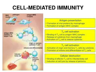
T cell activation and differentiation T helper 1 cells T helper 2 cells CD4 cells Regulatory T cells (Tregs) T helper 17 cells T cells CD8 cells (similar to CD4) NK T cells
Figure 8-39 CD4 + T cells Th1 cells activate macrophages to become highly bacteriocidal. Th1 cells coordinate the host response to intracellular pathogens, like tuberculosis. Th2 cells provide B cell help and promote antibody responses.
Cytotoxic CD8 T cells induce apoptosis of target cells Condensed chromatin, membrane vesicles are shed Condensed nucleus, no mitochondria, loss of cytoplasm.
Figure 8-31 Summary There are different classes of effector T-cells, specialized to deal with three classes of pathogen. Types of effector T cells produce distinct effector molecules. See notes for regulatory T cells and Th17 cells
Figure 8-10 Co-stimulation Activation of naïve T-cells requires two independent signals delivered by the same APC.
Figure 5-11 The human leukocyte antigen system (HLA)= major histocompatibility complex (MHC). A superlocus : large number of genes related to immune system. Chromosome 6, and encodes cell-surface antigen-presenting proteins and many other genes. Different classes have different functions: HLA class I antigens (that are produced from digested proteins A, B & C) present peptides from inside the cell (including viral peptides) that are broken down in the proteasomes. Foreign antigens attract killer T-cells (also called CD8 positive- or cytotoxic T-cells) that destroy cells. HLA class II antigens (DP,DM, DOA,DOB,DQ, & DR) present antigens from outside of the cell to T-lymphocytes. These particular antigens stimulate T-helper cells to multiply, and these T-helper cells then stimulate antibody-producing B-cells to produce antibodies to that specific antigen. Self-antigens are suppressed by HLA class III antigens encode components of the complement system. Other HLA roles: -important in disease defense -cause of organ transplant rejections -may protect against or fail to protect (if down regulated by an infection) against cancers -may mediate autoimmune disease (examples: type I diabetes, coeliac disease) Aside from the genes encoding the 6 major antigens, there are a large number of other genes, many involved in immune function, located on the HLA complex. Diversity of HLA in human population is one aspect of disease defense, and, as a result, the chance of two unrelated individuals having identical HLA molecules on all loci is very low. Historically, HLA genes were identified as a result of the ability to successfully transplant organs between HLA similar individuals.
Specific/adaptive immune responsesThe humoral immune response
Figure 7-1 B cell development
Figure 9-1 The humoral immune response is mediated by Ab molecules that are secreted by plasma cells (mature B cells). Ag that binds to B cell Ag receptor signals B cells and at the same time it is internalized and processed into peptides that activate armed T cells. Signals from the receptor and T cell induce the B cell to proliferate and differentiate into plasma cells. The functions of Ab. NB!
Figure 9-3 B cells and helper T cells must recognize the same molecular complex in order to interact.
Figure 9-11 part 3 of 3 Memory B cells then continue to recirculate through the B cell zones of secondary lymphoid tissues.
Figure 1-20 Memory is an important feature of the adaptive immune response.
Figure 1-16 Schematic representation of antibody structure.
Figure 9-19 Function and distribution of antibody IgM: pentamer, role in acute infection, activates complement +++, also neutralization of Ag and opsonization of pathogens. Occurs mainly in blood IgG: 4 isotypes, neutralization, opsonization and complement activation, crosses placenta, associated with more chronic forms of infection, diffuses into extravascular sites IgA: dimer, neutralization of antigen, complement activation, in mucosal secretions, role in ‘immune exclusion’ (keeps Ag outside and does not elicit major immune response as part of mucosal immune system. IgE: binds with Fc part to Fc receptors on mast cells, cross-binding leads to degranulation of mast cells (results in allergic manifestations), function in anti-parasite defenses. Occurs in submucosal sites. IgD: unknown function
Figure 9-22 Distribution of Ig isotypes in body (NB) Plasma Extracellular fluid Secretions across epithelia Mast cells beneath epithelia (skin, gut, respiratory epithelium
Figure 1-24 The functions of antibody (NB)
Figure 9-35 IgE cross-linking on mast cells leads to rapid release of inflammatory mediators.
A, B, AB, O blood groups Hemagglutination is used to type blood groups and match compatible donors and recipients for blood transfusion. Gut bacteria have antigens that are identical to blood-group antigens. Ab against these are only formed if the person does not have these antigens on their red cells. Before transfusion, the ABO blood group of the donor and recipient is determined by checking the agglutination pattern with anti-A and anti-B Ab. Before transfusion the serum of the recipient is tested for antibodies that agglutinate the red cells of the donor and vice versa= cross-match.
Donor blood groups Recipient blood groups