Download
1 / 48
590 likes | 1k Vues
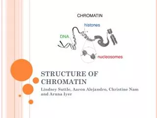
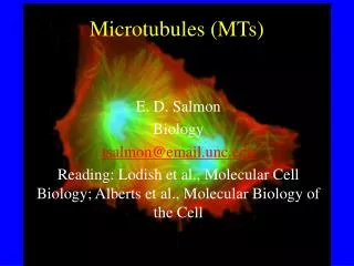
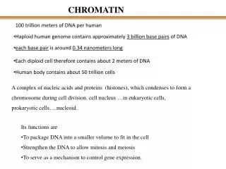
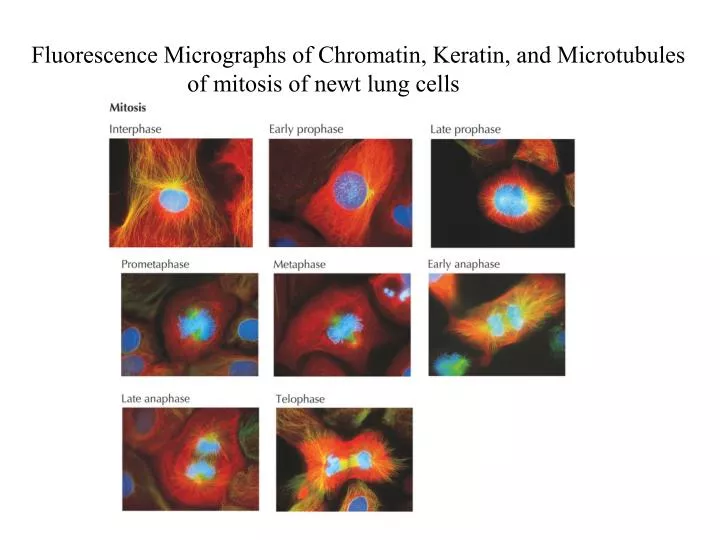
Fluorescence Micrographs of Chromatin, Keratin, and Microtubules of mitosis of newt lung cells.

E N D
Fluorescence Micrographs of Chromatin, Keratin, and Microtubules of mitosis of newt lung cells
Chapter 14 & 15Reading ListPages 560 – 569(On page 566, for controlled proteolysis, only a general understanding – no mechanism needed;Last two paragraphs on page 566 and pages 567-569 – a general understanding only- no need to memorize)Pages 642 – 646(For the extrinsic pathway on page 643 and other pathways on pages 644-646 – only a general understanding – no need to memorize)
Replication occurs during a defined period of the cell cycle
Identification of S Phase Cells by Incorporation of Radioactive Thymidine
Cell-fusion Experiments to Identify Cell cycle control proteins
Progesterone stimulates meiotic maturation of Xenopus oocytes in vitro
A diffusible factor in arrested Xenopus eggs promotes meiotic maturation in the absence of progesterone!Microinjection Assay:MPF Identified!
MPF Activity in Xenopus oocytes, eggs,and early embryos peaks as cells enter meiosis and mitosis.MPF activity was determined by the microinjection assay and quantitated by making dilutions of cell extracts
Biochemical Column Chromatography characterized MPF as a heterodimeric complex composed of a cyclin subunit and a CDK subunitThe challenge was to determine how these two subunits could induce mitosis?
Fluctuation of cyclin and MPF levels during the cell cycle of Xenopus
Experimental detection of cyclical synthesis and destruction of mitotic cyclin in sea urchin embryosA suspension of sea urchin eggs was synchronously fertilizedby the addition of sea urchin sperm, and radioactive 35 S Methionine was added. At 10 minute intervals beginning 16 minutes after fertilization, samples were taken for protein analysis by SDS-PAGE and for detection of cell cleavage by Microscopy
Experimental detection of cyclical synthesis and destruction of mitotic cyclin in sea urchin embryos
Regulation of mitotic cyclin levels in cycling Xenopus early embryonic cells
Recessive and dominant S.pombe cdc2 mutants have opposite phenotypes
Regulation of the kinase activity of S.pombe mitosis promoting factor (MPF)
Cdc-25 and Wee1 have opposing effects on S.pombe MPF activity
Strucural models of human CDK2 which is homologous to the S.pombe cyclin-dependent kinase (CDK)
Subcellular localization during the cell cycleHeLa cells injected with cyclin B1 linked to GFP G2 phase Prophase of Mitosis
P27: a Cdk inhibitor that arrests cell cycle progression p27-/- p27+/+ Thymus Gland
The survival of motor neurons depends on the size of the muscle target field they innervate : Experiments with Chick Embryos In the Early 1900’s!
Apoptosis: Genetic Studies in C.elegans947 non gonadal cells generated during development of the adult hermaphrodite form : 131 cells undergo cell deathcan be followed by Nomarski Interference Microscopy
Ced-3 gene in C.elegans is required for Cell death ced-3 mutation 131 cells survive
Genetic Studies in C.elegans Ced-3 mutation in C.elegans:All 131 cells survive!Ced-4 mutation in C. elegans:All 131 cells suviveCed-9mutation in C.elegans:All cells die during embryonic life – adult form never develops!Ced-9/Ced-3 Double Mutation in C.elegans:Cell Death Does Not Occur!Therefore, Ced-9 acts upstream of Ced-3 to suppress the apoptotic pathway
Confluence of genetic studies in Worms and Studies on Human Cancer cellsRegulator Proteins IdentifiedHuman bcl-2 gene cloned from human follicular lymphomas, since a mutated version of this gene was shown to act as a oncogene that promotes cell survival.The human Bcl2 protein was shown to be homologous to the worm CED-9 protein!A human bcl-2 transgene was able to block the extensive cell death found in ced-9 mutant worms! Thus both proteins act as regulators that suppress the apoptotic pathways.
Effector Proteins identifiedIn Worms, the ced-3 gene codes for the CED-3 protein which is a CaspaseEnzymeIn vertebrates, the gene ced-3 was shown to code for a caspase enzymeCaspases recognize and cleave short amino acid sequencs in many different target proteins in the cells
Cell Culture StudiesAdaptor Proteins identified that coordinate the action of regulatorsExpression of C.elegans CED-4 in a human kidney cell line induces rapid apoptosis!This can be blocked by coexpression of the negative regulator CED-9 (or mammalian Bcl-2) indicating that CED-9 opposes CED-4 action.CED-9 can interact directly with CED-4 Thus the pro-apoptotic function of CED-4 is directly suppressed by the anti-apoptotic function of CED-9.CED-4 also binds directly to the CED-3 caspase and promotes activation of its protease activity.Biochemical studies also indicate that CED-4 can simultaneosuly bind to both CED-9 and CED-3.Above results fit with the Genetics, which shows that the absence of CED-9 has no effect if CED-3 is also missing (ced-3/ced-9 double mutants have no cell death, like ced-3 mutants).
Overview of the evolutionarily conserved apoptotic pathway in C.elegans and vertebrates Regulator Adaptor Effector
For General KnowledgeProposed Intracellular Pathways leading to cell death by apoptosis or to trophic factor-mediated cell survival in mammalian cells.
Study Model in your Text BookChapter 15 Pages 643-646No need to memorize ModelOnly Understand it!