Download
1 / 80
820 likes | 1.09k Vues
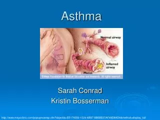
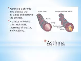
ASTHMA. What is Asthma. A Chronic disease of the airways that may cause: Wheezing Breathlessness Chest tightness Nighttime or early morning coughing.

E N D
What is Asthma • A Chronic disease of the airways that may cause: • Wheezing • Breathlessness • Chest tightness • Nighttime or early morning coughing
The bronchospasm characteristic of the acute asthmatic attack is typically reversible. It improves spontaneously or within minutes to hours of treatment
Asthma can exist by itself or coexist with chronic bronchitis, emphysema, or bronchiectasis
Symptoms/Chief Complaint • Progressive dyspnea • Cough • Chest tightness • Wheezing/coughing
The rapidly reversible airflow obstruction of asthma is mainly due to bronchial smooth muscle contraction
Focus of Therapy • Pharmacologic manipulation of airway smooth muscle • Do not overlook physiologic impairment caused by mucous production and mucosal edema • Bronchospasm can be reversed in minutes • Airflow obstruction due to mucous plugging and inflammatory changes in bronchial walls may not resolve for days/weeks - • may lead to atelectasis, infectious bronchitis, pneumonitis
Asthma Triggers • Immunologic reaction • Viral respiratory/sinus infections • change in temperature/humidity • Drugs/Chemicals - • aspirin, NSAIDS • Exercise • GE reflux • Laughing/coughing • Environmental factors - • strong odors, pollutants, dust, fumes
Patient Exam • Wheezing • may be audible w/o stethoscope • Use of accessory muscles of inspiration • diaphragmatic fatigue • Paradoxical respirations • - reflect impending ventilatory failure • Altered mental status - • lethargy, exhaustion, agitation, confusion
Patient Exam • Hyperrsonance to percussion • decreased intensity of breath sounds • prolongation of expiratory phase w or w/o wheezing
Patient Exam • The intensity of the wheeze may not correlate with the severity of airflow obstruction • “quiet chest” - very severe airflow obstruction
Risk factors for death from asthma • Past history of sudden severe exacerbations • Prior intubation for asthma • Prior admission for asthma to an intensive care unit • Two or more hospitalizations for asthma in the past year • Three or more emergency care visits for asthma in the past year • Hospitalization or emergency care visit for asthma within the past month
Risk factors cont.. • Use of more than two canisters per month of inhaled short-acting 2-agonist • Current use of systemic corticosteroids or recent withdrawal from systemic corticosteroids • Difficulty perceiving airflow obstruction or its severity • Comorbidity, as from cardiovascular diseases or chronic obstructive pulmonary disease • Serious psychiatric illness or psychosocial problems • Low socioeconomic status in urban residents • Illicit drug use
Asthma Treatment • Nebulized B-adrenergic drugs • Corticosteroids • Nebulized anticholinergics • Magnesium sulfate • Oxygen • Long acting beta-agonists • Inhaled steroids
Managing Asthma: • Indications of a severe attack: • Breathless at rest • hunched forward • talking in words rather than sentences • Agitated • Peak flow rate less than 60% of normal
Treatment Goals of Severe Asthma • Improve airway function rapidly • Avoid hypoxemia • Prevent respiratory failure and death
Mild Moderate Severe walking talking at rest Can lie down Prefers sitting upright Sentences Phrases Words May be agitated Usually agitated Symptoms Breathlessness Position Talks in Alertness Classifying Severity of Asthma Exacerbations
Classifying Severity of Asthma Exacerbations Signs Mild Moderate Severe
Classifying Severity of Asthma Exacerbations Functional assessmentMild Moderate Severe
80% 50–80% <50% or response lasts<2h Normal >60 mm Hg <60 mm Hg: possible cyanosis <42 mm Hg <42 mm Hg > 42 mm Hg: possible respiratory failure >95 %91–95% <91% Peak expiratory flow % predicted or % personal best PaO2 (on air) PaCO2 SaO2% (on air) at sea level Classifying Severity of Asthma Exacerbations Functional assessmentMild Moderate Severe
Respiratory Arrest Imminent • Drowsy or confused • Paradoxical thoracoabdominal movement • Absent Wheeze • Bradycardia • Absence Pulsus paradoxus suggests respiratory muscle fatigue
Asthma Mimickers • Congestive heart failure ("cardiac asthma") • Upper airway obstruction • Aspiration of foreign body or gastric acid • Bronchogenic carcinoma with endobronchial obstruction • Metastatic carcinoma with lymphangitic metastasis • Sarcoidosis with endobronchial obstruction • Vocal cord dysfunction • Multiple pulmonary emboli (rare)
treatment of acute asthma Goal in the ED • reverse airflow obstruction rapidly by repetitive or continuous administration of inhaled 2-agonists • ensure adequate oxygenation • relieve inflammation
Initial Assessment • History • physical examination (auscultation use of accessory muscles, heart rate, respiratory rate) • PEFR or FEV • oxygen saturation • other tests as indicated
Diagnosis • Bedside spirometry • rapid, objective assessment ,guide to the effectiveness of therapy. • The forced expiratory volume in 1 s (FEV1) • peak expiratory flow rate (PEFR) • Sequential measurements • management decisions
Pulse oximetry • assessing oxygenation and monitoring oxygen saturation during treatment. • ABG is not indicated in most patients with mild to moderate asthma exacerbation
ABG assess for hypoventilation with carbon dioxide retention and respiratory acidosis • clinical evidence of severe attacks • PEFR or FEV1 of less than 25 percent predicted • With acute attacks, ventilation is stimulated, resulting in a decrease in partial pressure of carbon dioxide (PaCO2) • normal or slightly elevated PaCO2 (e.g., 42 mm Hg) indicates extreme airway obstruction and fatigue and may herald the onset of acute ventilatory failure
radiography • clinical indication of a complication • pneumothorax, pneumomediastinum, pneumonia, or other medical concern • one-third of asthma exacerbations requiring admission, will demonstrate an abnormality on chest radiograph
CBC • not indicated • modest leukocytosis secondary to administration of B -agonist therapy or corticosteroid treatment • In patients taking theophylline before ED presentation, a serum theophylline level
ECG • Routine electrocardiogram is unnecessary right ventricular strain, abnormal P waves, or nonspecific ST- and T-wave abnormalities, which resolve with treatment Older patients, especially those with coexisting heart disease, should have cardiac monitoring during treatment
Impending or Actual RespiratoryArrest • Intubation and mechanical ventilation with 100% 02 • Nebulized B2 agonist and anticholinergic • Intravenous steroid • Admit to ICU
Repeat Assessment • Symptoms. • physical examination. • PEFR. • 02 saturation. • other test as needed
medications are used in the treatment of acute asthma • adrenergic agonists • anticholinergics • glucocorticoids • Magnesium, heliox (mixture of helium and oxygen), and ketamine may be considered when the aforementioned medications fail to relieve bronchospasm. • Mast cell-stabilizing agents, methylxanthines, and leukotriene modifiers are currently reserved for maintenance therapy only
Adrenergic Agents • Adrenergic receptors • Stimulation of B 1-receptors increases rate and force of cardiac contraction and decreases small intestine motility and tone • B2-adrenergic stimulation promotes bronchodilation, vasodilation, uterine relaxation, and skeletal muscle tremor
Adrenergic Agents • stimulation of the enzyme adenyl cyclase, which converts intracellular adenosine triphosphate into cyclic adenosine monophosphate • enhances the binding of intracellular calcium to cell membranes, reducing the myoplasmic calcium concentration, and results in relaxation of bronchial smooth muscle • inhibit mediator release and promote mucociliary clearance.
side effect of B-adrenergic drugs • skeletal muscle tremor (most common) • nervousness, anxiety, • insomnia, headache, • hyperglycemia, • palpitations, tachycardia, and hypertension • potential cardiotoxicity(combination with theophylline not significant problems) • Arrhythmias and evidence of myocardial ischemia( rare)
Inhaled short-acting B-2 agonists Albuterol • Nebulizer solution (5 mg/mL) • 2.5–5.0 mg every 20 min for 3 doses • then 2.5–10 mg every 1–4 h as needed or 10–15 mg per h continuously • Only selective B-2 agonists are recommended • for optimal delivery, dilute aerosols to minimum of 4 mL at gas flow of 6–8 L per min
Albuterol • MDI (90 g/puff) • 4–8 puffs every 20 min up to 4 h • then every 1–4 h as needed • As effective as nebulized therapy if patient is able to coordinate inhalation maneuver; use spacer/holding chamber
Inhaled short-acting B-2 agonists • Bitolterol • Nebulizer solution (2 mg/mL) • MDI (370 macg/puff) • Pirbuterol • MDI (200 g/puff)