Download
1 / 42
530 likes | 1.12k Vues
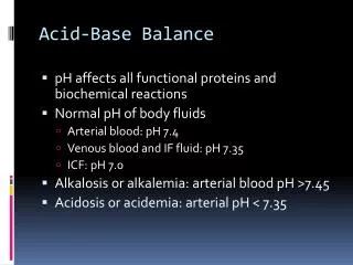
Acid-Base Balance. Acid-Base Balance. Normal pH of body fluids Arterial blood is 7.4 Venous blood and interstitial fluid is 7.35 Intracellular fluid is 7.0 Alkalosis or alkalemia – arterial blood pH rises above 7.45 Acidosis or acidemia – arterial pH drops below 7.35. Figure 24.5 2.

E N D
Acid-Base Balance • Normal pH of body fluids • Arterial blood is 7.4 • Venous blood and interstitial fluid is 7.35 • Intracellular fluid is 7.0 • Alkalosis or alkalemia– arterial blood pH rises above 7.45 • Acidosis or acidemia– arterial pH drops below 7.35
Figure 24.5 2 The narrow range of normal pH of the ECF, and the conditions that result from pH shifts outside the normal range The pH of the ECF(extracellular fluid)normally ranges from7.35 to 7.45. When the pH of plasma falls below7.5, acidemia exists. Thephysiological state that results iscalled acidosis. When the pH of plasma risesabove 7.45, alkalemia exists.The physiological state thatresults is called alkalosis. Extremelyacidic Extremelybasic pH Severe acidosis (pH below 7.0) can be deadlybecause (1) central nervous system functiondeteriorates, and the individual may becomecomatose; (2) cardiac contractions grow weak andirregular, and signs and symptoms of heart failuremay develop; and (3) peripheral vasodilationproduces a dramatic drop in blood pressure,potentially producing circulatory collapse. Severe alkalosis is alsodangerous, but serious casesare relatively rare.
Acid–Base Balance - Hydrogen Ions (H+) • Are gained • At digestive tract • Through cellular metabolic activities • Are eliminated • At kidneys and in urine • Must be neutralized to avoid tissue damage • Acids produced in normal metabolic activity • Are temporarily neutralized by buffers in body fluids
pH and enzyme function • Hydrogen ion concentration has a widespread effect on the function of the body's enzyme systems. • The hydrogen ion is highly reactive and will combine with bases or negatively charged ions at very low concentrations. • Proteins contain many negatively charged and basic groups within their structure. • Thus, a change in pH will alter the degree ionization of a protein, which may in turn affect its functioning. • At more extreme hydrogen ion concentrations a protein's structure may be completely disrupted (the protein is then said to be denatured).
Sources of Hydrogen Ions • Most hydrogen ions originate from cellular metabolism • Breakdown of phosphorus-containing proteins releases phosphoric acid into the ECF • Anaerobic respiration of glucose produces lactic acid • Fat metabolism yields organic acids and ketone bodies • Transporting carbon dioxide as bicarbonate releases hydrogen ions
CO2 + H20 H2CO3 HCO3- + H+ Types of acids in the body • Volatile acidcomes from carbohydrate and fat metabolism • Can leave solution and enter the atmosphere (e.g. carbonic acid – H2CO3) • Breaks in the lungs to carbon dioxide and water • In the tissues CO2 reacts with water to form carbonic acid, which dissociate to give hydrogen ions and bicarbonate ions • This reaction occurs spontaneously, but happens faster with the presence of carbonic anhydrase (CA) • PCO2 and pH are inversely related
Types of acids in the body • Fixed acids • Acids that do not leave solution (e.g. sulfuric and phosphoric acids – produced during catabolism of amino acids) • Eliminated by the kidneys • Organic acids • by-products of anerobic metabolism such as lactic acid, ketone bodies
Buffers • Buffers- compound that limits the change in hydrogen ion concentration (and so pH) when hydrogen ions are added or removed from the solution. http://www.nda.ox.ac.uk/wfsa/html/u13/u1312f03.htm
Buffer systems • Two types of buffer in the body • Chemical buffers • Bicarbonate, phosphate and protein systems • Substance that binds H+ and remove it from the solution if its concentration rises or release it if concentration decreases • Fast reaction within seconds • Physiological • respiratory (fast reaction – few minutes) or urinary (slow reaction – hours to days) • Regulates pH by controlling the body’s output of bases, acids or CO2
Acid-Base Balance • Hydrogen ion and pH balance in the body CO2 (+ H2O)Lactic acidKetoacids Fatty acidsAmino acids H+ input Plasma pH7.38–7.42 Buffers:• HCO3– in extracellular fluid• Proteins, hemoglobin, phosphates in cells• Phosphates, ammonia in urine CO2 (+ H2O) H+ output H+ Figure 20-18
Figure 24 Section 2 1 The major factors involved in the maintenanceof acid-base balance The respiratory systemplays a key role byeliminatingcarbon dioxide. The kidneys play a majorrole by secretinghydrogen ions into the urine and generatingbuffers that enter thebloodstream. The rate ofexcretion rises and fallsas needed to maintainnormal plasma pH. As a result, the normal pH ofurine varies widely butaverages 6.0—slightlyacidic. Active tissuescontinuously generatecarbon dioxide, which insolution forms carbonicacid. Additional acids,such as lactic acid, areproduced in the course ofnormal metabolicoperations. Normalplasma pH(7.35–7.45) Tissue cells Buffer Systems Buffer systems cantemporarily store Hand thereby provideshort-term pHstability.
Chemical Buffer Systems • Three major chemical buffer systems • Bicarbonate buffer system • Phosphate buffer system • Protein buffer system • Any drifts in pH are resisted by the entire chemical buffering system
Bicarbonate Buffer System • A mixture of carbonic acid (H2CO3) and its salt, sodium bicarbonate (NaHCO3) (potassium or magnesium bicarbonates work as well) • If strong acid is added: • Hydrogen ions released combine with the bicarbonate ions and form carbonic acid (a weak acid) • The pH of the solution decreases only slightly • If strong base is added: • It reacts with the carbonic acid to form sodium bicarbonate (a weak base) • The pH of the solution rises only slightly • This system is the only important ECF buffer CO2 + H2O H2CO3 H+ + HCO3¯
Figure 27-9 The Basic Relationship between PCO2 and Plasma pH H2O CO2 H2CO3 H HCO3 PCO2 40–45 mm Hg HOMEOSTASIS If PCO2rises When carbon dioxide levels rise, more carbonic acid forms, additional hydrogen ions and bicarbonate ions are released, and the pH goes down. PCO2 pH
Figure 27-9 The Basic Relationship between PCO2 and Plasma pH H HCO3 H2CO3 H2O CO2 pH 7.35–7.45 HOMEOSTASIS If PCO2falls When the PCO2 falls, the reaction runs in reverse, and carbonic acid dissociates into carbon dioxide and water. This removes H ions from solution and increases the pH. pH PCO2
Phosphate Buffer System • Nearly identical to the bicarbonate system • Its components are: • Sodium salts of dihydrogen phosphate (H2PO4¯), a weak acid • Monohydrogen phosphate (HPO42¯), a weak base • This system is an effective buffer in urine and intracellular fluid
Protein Buffer System • Plasma and intracellular proteins are the body’s most plentiful and powerful buffers • Some amino acids of proteins have: • Free organic acid groups (weak acids) • Groups that act as weak bases (e.g., amino groups) • Amphoteric molecules are protein molecules that can function as both a weak acid and a weak base
Figure 27-11 The Role of Amino Acids in Protein Buffer Systems Neutral pH If pH rises If pH falls Amino acid In alkaline medium, amino acid acts as an acid and releases H In acidic medium, amino acid acts as a base and absorbs H
Buffer Systems in Body Fluids Figure 27.7
Physiological Buffer Systems – respiratory system • The respiratory system regulation of acid-base balance is a physiological buffering system • The respiratory buffering system takes care of volatile acids – by-products of glucose and fat metabolism CO2 + H2O H2CO3 H+ + HCO3¯
Physiological Buffer Systems – respiratory system • During carbon dioxide unloading, hydrogen ions are incorporated into water • When hypercapnia or rising plasma H+ occurs: • Deeper and more rapid breathing expels more carbon dioxide • Hydrogen ion concentration is reduced • Alkalosis causes slower, more shallow breathing, causing H+ to increase
pH Disturbances • The reflex pathway for respiratory compensation of metabolic acidosis Plasma H+( pH) PlasmaPCO2 by Law of Mass Action Carotid and aorticchemoreceptors Centralchemoreceptors Sensory neuron Interneuron Respiratorycontrol centersin themedulla Negative feedback Negative feedback Action potentials in somaticmotor neurons Muscles of ventilation Rate and depth of breathing PlasmaPCO2 Plasma H+( pH) by Law of Mass Action Figure 20-19
Physiological Buffer Systems – kidneys • Chemical buffers can tie up excess acids or bases, but they cannot eliminate them from the body • The lungs can eliminate carbonic acid by eliminating carbon dioxide • Only the kidneys can excrete the body of metabolic acids (phosphoric, uric, and lactic acids and ketones) and prevent metabolic acidosis • The ultimate acid-base regulatory organs are the kidneys
Physiological Buffer Systems – kidneys • The kidney takes care of the non-volatile acid products • By-products of protein metabolism and anaerobic respiration • The kidneys must prevent the loss of bicarbonate ions (re-absorb) that is being constantly filtered from the blood. • Both tasks are accomplished by secretion of hydrogen ions • Only about 10% of the hydrogen ions secreted will be excreted • As a result of the H+ excretion the urine is usually acidic
Renal compensation when pH is low • When H+ or PCO2 in plasma is high – acidosis • The kidneys will: • Secrete H+ in nephron and excretion will increase • All the filtered HCO3- will be reabsorbed • Produce HCO3- to increase its blood levels
Renal Compensation • Hydrogen Ions • Are secreted into tubular fluid along: • Proximal convoluted tubule (PCT) • Distal convoluted tubule (DCT) • Collecting system
Renal Compensation • The ability to eliminate large numbers of H+ in a normal volume of urine depends on the presence of buffers in urine • Major Buffers in Urine • Glomerular filtration provides components of: • Carbonic acid–bicarbonate buffer system • Phosphate buffer system • Tubule cells of PCT • Generate ammonia
Reabsorption of Bicarbonate • In a person with normal acid-base balance all the HCO3- in the tubular fluid is consumed by neutralizing H+ - no HCO3- in the urine • HCO3- molecules are filtered by the glomerulus and than reabsorbed and appear in the peritubular capillary (most in the PCT). • The re-absorption is not direct – the luminar surface of the tubular cells can not absorb HCO3- • The kidney cells can also generate new HCO3- if needed
Renal compensation when pH is high • When H+ or PCO2 in plasma is low – alkalosis • The kidneys will: • Inhibit secretion of H+ in nephron and excretion will decrease • Reduced HCO3- reabsorption; will appear in urine and level in plasma decrease
Acid–Base Balance Disturbances • Respiratory Acid–Base Disorders • Result from imbalance between: • CO2 generation in peripheral tissues • CO2 excretion at lungs • Cause abnormal CO2 levels in ECF • Metabolic Acid–Base Disorders • Result from: • Generation of organic or fixed acids • Conditions affecting HCO3- concentration in ECF
Respiratory Acidosis and Alkalosis • Result from failure of the respiratory system to balance pH • PCO2 is the most important indicator of respiratory inadequacy • PCO2 levels • Normal PCO2 fluctuates between 35 and 45 mm Hg • Values above 45 mm Hg signal respiratory acidosis • Values below 35 mm Hg indicate respiratory alkalosis
Respiratory Acidosis and Alkalosis • Respiratory acidosis is the most common cause of acid-base imbalance • Occurs when a person breathes shallowly, or gas exchange is slowed down by diseases such as pneumonia, cystic fibrosis, or emphysema • Respiratory alkalosis is a common result of hyperventilation
Metabolic Acidosis • Metabolic acidosis is the second most common cause of acid-base imbalance • Can be a result of: • Failure of the kidney to excrete metabolic acids • Renal acidosis is either the inability of kidney to excrete H+ or to re- absorb bicarbonate ion • Diarrhea – most common reason of metabolic acidosis • Loss of large amounts of sodium bicarbonate in the feces (which is normal component of the feces) • Diabetes mellitus – results in breakdown of fat that releases acids • Ingestion of acids • Acetylsalicylic acid (aspirin) • Methyl alcohol (forms acid when metabolized)
Metabolic Alkalosis • Is caused by elevated HCO3–concentrations • Bicarbonate ions interact with H+ in solution • Forming H2CO3 • Reduced H+ causes alkalosis • Typical causes are: • Vomiting of the acid contents of the stomach • Intake of excess base (e.g., from antacids) • Constipation, in which excessive bicarbonate is reabsorbed
Acid-base imbalances • To be able to assess the type of imbalance: • 1. look at the pH – to decide acidosis/alkalosis • 2. look at PCO2 and HCO3- to decide respiratory/metabolic • If PCO2 causes the acidosis/alkalosis – it is respiratory • If HCO3- causes the acidosis/alkalosis – it is metabolic
To determine compensation: • Uncompensated= abnormal pH and change in one blood parameter • Partially compensated= all 3 values of pH, HCO3-, CO2 are abnormal • Fully compensated= pH is normal, both HCO3- and CO2 are abnormal • Corrected= all parameters are normal
The response to acidosis caused by the addition of H Addition of H Start CARBONIC ACID-BICARBONATE BUFFER SYSTEM BICARBONATE RESERVE H2CO3 HCO3 Na CO2 CO2 H2O HCO3 NaHCO3 H (bicarbonate ion) (carbonic acid) (sodium bicarbonate) Lungs Generation of HCO3 Other buffer systems absorb H KIDNEYS Respiratory Response to Acidosis Renal Response to Acidosis Increased respiratory rate lowers PCO2, effectively converting carbonic acid molecules to water. Kidney tubules respond by (1) secreting H ions, (2) removing CO2, and (3) reabsorbing HCO3 to help replenish the bicarbonate reserve. Secretion of H
The response to alkalosis caused by the removal of H Removal of H Start CARBONIC ACID-BICARBONATE BUFFER SYSTEM BICARBONATE RESERVE Lungs HCO3 Na CO2 H2O H2CO3 HCO3 NaHCO3 H (bicarbonate ion) (sodium bicarbonate) (carbonic acid) Generation of H Other buffer systems release H Respiratory Response to Alkalosis KIDNEYS Decreased respiratory rate elevates PCO2, effectively converting CO2 molecules to carbonic acid. Renal Response to Alkalosis Kidney tubules respond by conserving H ions and secreting HCO3. Secretion of HCO3