Download
1 / 30
310 likes | 442 Vues
Integumenatry System. Ch 5. The Skin as an Organ. Function. Protection- mechanical, chemical, bacterial Body temperature regulation Prevent water loss Metabolic- synthesize vitamin D Cutaneous Sensation Blood reservoir Excretion Prevent UV damage. The Skin.

E N D
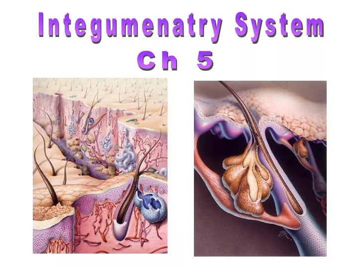
Integumenatry System Ch 5
Function • Protection- mechanical, chemical, bacterial • Body temperature regulation • Prevent water loss • Metabolic- synthesize vitamin D • Cutaneous Sensation • Blood reservoir • Excretion • Prevent UV damage
The Skin An organ, cells constantly dying and being replaced • Facts: • Weighs 9-11 lbs • s.a. = 1.5-2m2 • 1 cm2 has 70 cm blood vessels, 55 cm • nerves, 100 sweat glands, 15 oil glands, 230 sensory receptors • New skin produced in 25-45 days
Cells of the Epidermis • Keratinocytes (90%)- waterproofs & protects skin, nails, hair, stratum corneum • Melanocytes (8%)- produce melanin • Merkel Cells- slow mechanoreceptors • Langerhans’ Cells- immunological defense
Layers of the Epidermis • Stratum Corneum • Stratum Lucidum • Stratum Granulosum • Stratum Spinosum • Stratum Basale- (Germinativum)
The Dermis Dermis
Layers of the Dermis papillary dermis reticular dermis
Components of the Dermis a. Cellular Fibroblasts (synthesize collagen, elastin, and reticulin), histiocytes, endothelial cells, perivascular macrophages and dendritic cells, mast cells, smooth muscle, and cells of peripheral nerves and their end-organ receptors. b. Fibrous Collagen & reticulin - provide tensile strength Elastic fibers- provide for restoration of shape after a deformation c. Ground substance glycosaminoglycans: hyaluronic acid, chondroitin sulfate, and dermatan sulfate.
The Hypodermis Hypodermis This layer contains adipose tissue and serves to attach the dermis to its underlying tissues.
Skin Color Some variations in human skin color (Sub-Saharan African, Indian, Southern European, and Northwest European) Skin color due to: Melanin, Carotene & Hemoglobin • Melanin Pigments: • Eumelanin: • Phaeomelanin • > Eumelanin:Phaeomalanin- darker skin and hair color; • < Eumelanin:Phaeomalanin- lighter skin and hair color
Skin Color Human complexions are generally classified into six skin types: I -light skinned, burns easily, never tans II - light skinned, burns easily, tans some III - light skinned, burns occasionally, tans well IV - light skinned, tans well, rarely burns V - brown skinned (Asian, Indo-Asian, Chinese, Japanese), tans well, burns rarely, can sunburn after prolonged exposure to UVR VI - black skinned (Afro-Caribbean), deeply pigmented, can burn after prolonged exposure to UVR 25% US pop
Skin Color Conditions • Cyanotic • Jaundice • Erythema • Pallor
Skin/Hair Color: Pigmentation • Pigmentation levels usually increase with age. • - exception: premature graying • Normal pigmentation may be altered by genetic defects or by acquired diseases. • -Hyperpigmentation- age spots • -Hypopigmentation- vitiligo, • albinism
Skin/Hair Color: Pigmentation • External agents can also alter skin color. • lightening agents • carotene • dyes • Some internal compounds--such as the byproducts of hemoglobin metabolism--may color the skin. Sunless tanning
Skin Cancer • Malignant melanoma • 2% of all cancers • Risks: • Skin type • Sun exposure • Family history • Age • Immunological status Normal mole Melanoma • A= asymmetry • B= border • C= color • D= diameter
Skin Appendages Sweat Glands • Eccrine (merocrine) glands- sweat • Apocrine glands- axillary & anogenital areas • Ceruminous glands- ears canal • Mammary glands- female reproductive glands Sweat glands Ceruminous glands
Skin Appendages Sebaceous
Skin Appendages Hair Hair shaft Sebaceous gland Hair root Hair bulb in follicle
Skin Appendages Nail
Burns 1st Degree: epidermal damage Ex. sunburn 2nd Degree: epidermis & upper dermis Ex. blisters 3rd Degree: entire thickness of skin
Burns Skin replacement: http://www.youtube.com/watch?v=eXO_ApjKPaI
Tissue Repair • Blood vessels dilate • WBC & clotting agents released • Scab forms
Tissue Repair • Granulation tissue forms • Capillary beds invade clot • Clean up begins
Tissue Repair • Scar area has contracted • Epithelium regeneration begins
INQUIRY • In which lay of skin are blood vessels located? • Where does epithelium regeneration begin? • What color is a persons skin if they are cyanotic? • List the layer of the epidermis in order from top to bottom. • What is the primary tissue of the hypodermis?