Download
1 / 65
650 likes | 816 Vues
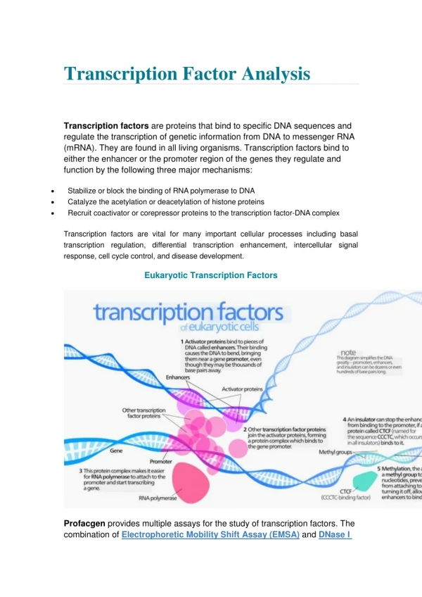
Location analysis of transcription factor binding sites. Guy Naamati Andrei Grodzovky. A brief history. What about today?. Two weeks ago Masha and Michal told us about gene expression and gene clusters. Last week, Lior and Ofer told us about tfbs, and how to identify them. Today!.

E N D
Location analysis of transcription factor binding sites Guy Naamati Andrei Grodzovky
A brief history • What about today? • Two weeks ago Masha and Michal told us about gene expression and gene clusters. • Last week, Lior and Ofer told us about tfbs, and how to identify them.
Today! • A revolutionary new method that identifies where and when in the genome a binding factor actually binds! • We will talk about the method that reveals the genome wide localization, and provide several important examples from the world of yeast cells.
Motivation no 1 • What can location analysis give us that micro-arrays alone can’t? • Micro-arrays identifies changes in mRNA levels, but can not distinguish direct from indirect effects.
Motivation no 2 • What advantage does localization have over try to identify the binding site? • Right! We don’t have to handle many case in which it “looks” like we identified binding site, but in vivo it’s not.
The Method • Developed by the group of Richard A. Young in Cambridge. • A combination of location and expression profile. • Allows protein-DNA interactions to be monitored across the entire yeast genome.
The Method • A modified ChIP, combined with micro-array analysis. • DNA was taken from a cell, and broken with sound waves (sonication). • Proteins of interest where tagged with myc. • Fragments cross linked to those proteins were enriched by immunoprecipitation (IP).
What now? • Cross links were reversed, and the enriched DNA was amplified and labeled (Cy5). • Cy5 labeled DNA was hybridized to a micro-array, together with non-enriched DNA labeled with Cy3. • Gene expression was also analyzed. • Three independent experiments, for accuracy.
Handling noise • A single-array error method was used.
How accurate is it? • This method can identify factors binding to DNA, but cannot recognize the exact location of the binding site. Why? • The sonication breaks the DNA into fragments 500-1000 bases long. Not very specific.
Testing if it works • Used to identify sites bound by Gal4 in the yeast genome. • Found seven genes previously reported to be regulated by Gal4. • In addition, 3 more genes were found!
An important reminder • The consensus binding site for Gal4 was found in many places in the gene where Gal4 did not bind. Why is that? • Previous studies of Gal4 have suggested that chromatin structure also has a big role.
The next investigation • Ste12 functions in the response of haploid yeast to mating pheromones. • More than 200 genes are activated in a Ste12 dependent fashion. Which are directly regulated? • By this method, only 29!
What’s next? • This method can identify the global set of genes that are regulated directly in vivo. • Gives us accurate information about where and when transcription factors bind. • Opens a new pathway into regulation analysis…
Just as there are networks of metabolic pathways… There are networks of regulator-gene interactions
But the network consists of building blocks : Those are… Network Motifs
How we identify them ? • Using genome wide location analysis • Identification of a set of promoter regions that are bound by specific regulators allowed us to predict sequence motifs that are bound by these regulators
Offers the potential to produce bistable systems that can switch between two alternative states.
Provides a form of multi step ultra sensitivity as small changes in the level of activity of the master regulator at the top of the loop might be amplified at the ultimate target.
Single Input Motif Single Input Motif Single-input motifs are potentially useful for coordinating a discrete unit of biological function, such as a set of genes that code for the subunits of a biosynthetic apparatus or enzymes of a metabolic pathway.
This motif offers the potential for coordinating gene expression across a wide variety of growth conditions.
The chain represents the simplest circuit logic for ordering transcriptional events in a temporal sequence.
Single Input Motif Single Input Motif Example FHL1 – Ribosomal proteins regulator. Genome wide location analysis Forms a single input regulatory motif consisting of essentially all ribosomal protein genes
Assembling motifs into network structures • An algorithm based on genome wide location data and expression data from over 500 experiments was developed in order to identify group of genes that are both coordinately bound and expressed.
Network assembly algorithm • 1-Define a set of genes G bound by a set of regulators S. • 2- Find a subset of G with a similar expression pattern. • 3- Go over the genes in G and drop genes with a significantly different expression pattern. • 4- Scan the remaining genome for genes with similar expression profile and check if they’re bound by factors from S.
What have we got ? • The resulting sets of genes and regulators are multi input motifs. • But they are refined for common expression MIM-CE
MIM-CE’s: What are they good for ? • Using MIM-CE’s the yeasts cell cycle networks was constructed using an automated method, without prior knowledge of the regulators that control transcription.
The process • Check for MIM-CE’s significantly enriched in genes whose expression oscillates during the cell cycle. • Align MIM-CE’s around the cell cycle on the basis of peak expression of the genes in the MIM-CE.
The outcome Yeasts cell cycle transcriptional regulatory network.
Features of the network model: • Correlation of the computational positioning of regulators with previous studies. • Regulators whose function was not known before could be positioned in the network on the basis of direct binding data. • Third, and most important, reconstruction of the regulatory architecture was automatic and required no prior knowledge of the regulators that control transcription during the cell cycle.
Serial Regulation of Transcriptional Regulatorsin the Yeast Cell CycleSimon et al.
Many transcriptional regulatory networks in yeast… Why the cell cycle network ?
Cyclins regulate the cell cycle Regulation of the cell cycle clock is effected through activity of the cyclin-dependent kinase (CDK) family of protein kinases.
But who regulate the regulators ? • Nine transcriptional regulators were identified
The method • Using genome wide location analysis to identify the binding sites for each of the factors in vivo.
The results ChIP Micro Array
These results confirm the stage specific regulation of gene expression by those factors.
The results also confirm that genes encoding several of the cell cycle transcriptional regulators are themselves bound by other cell cycle regulators In this way a full regulatory network is formed.
And of course the cell cycle regulators Cyclin’s/CDK’s are also regulated by those factors.
Functional redundancy • Each of the factors binds a critical cell cycle gene. • Deletion mutants with one of the factors deleted survive… • Why ?