Download
1 / 31
330 likes | 631 Vues
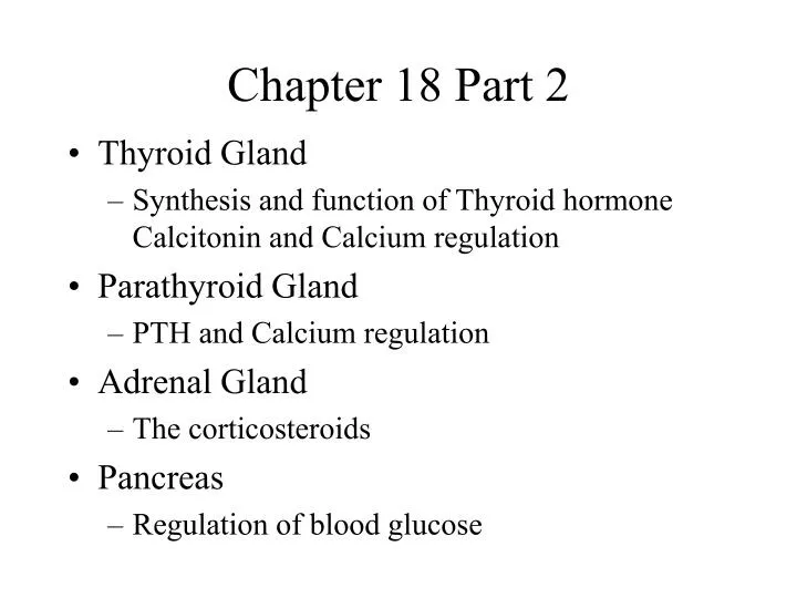
Chapter 18 Part 2. Thyroid Gland Synthesis and function of Thyroid hormone Calcitonin and Calcium regulation Parathyroid Gland PTH and Calcium regulation Adrenal Gland The corticosteroids Pancreas Regulation of blood glucose. Thyroid Gland. Hyoid Bone. lobe of thyroid. lobe of thyroid.

E N D
Chapter 18 Part 2 • Thyroid Gland • Synthesis and function of Thyroid hormone Calcitonin and Calcium regulation • Parathyroid Gland • PTH and Calcium regulation • Adrenal Gland • The corticosteroids • Pancreas • Regulation of blood glucose
Thyroid Gland Hyoid Bone lobe of thyroid lobe of thyroid • Covers anterior surface of trachea • 2 lobes with isthmus Isthmus Trachea
Thyroid gland • Tissue consists of follicles • Follicles are hollow spheres lined with epithelial cells (follicle cells) • Follicular cells take up Iodine from circulation and produce thryoglobulin (the precursor to thyroid hormone) • Thyroglobulin is stored in the colloid of follicle • C cells produce calcitonin (CT)
Thyroid Gland An elusive C-cell is indicated Here thyroglobulin, a glycoprotein, is stained hot pink
Synthesis of thyroid hormone • A funky, multistep process • Iodine selectively pumped into the membrane of follicular cells • Iodine bound to tyrosine molecules, forming thyroid hormone, incorporated into thyroglobulin • Thyroglobulin is stored in follicle • Upon TSH stimulation, thyroglobulin is endocytosed back into follicle cell, diffuses across cell, and released into bloodstream
T4 versus T3 • What is the difference? • The thyroid releases 90% T4, 10% T3 • T3 is the active form!
Functions of Thyroid Hormone • Actively transported into all cells of body • Binds mitochondria, increases rate of mitochondrial ATP production • Binds nuclear receptors and increases transcription of Na+/K+ ATPase • Also activates genes that code for enzymes involved in glycolysis and ATP production • PUNCHLINE: Thyroid hormone increases basal metabolic rate
Misregulation and Goiters • TSH causes thyroid hormone release AND growth of thyroid tissue
C cells and Calcitonin • C cells respond directly to high levels of Ca2+ in body fluids • Release Calcitonin (CT), works to reduce Ca2+ concentration in body fluids • How??
Calcitonin and Ca2+ regulation • Inhibition of osteoclasts (the bone breaker-downer cells) • Stimulation of Ca2+ excretion at kidneys CT osteoclast
Parathyroid Glands • Located on the posterior aspect of the thyroid gland • Chief cells produce and release parathyroid hormone (PTH) directly in response to low circulating Ca2+ levels • PTH works to increase Ca2+ levels
Parathyroid Glands • Stimulates osteoclasts • Inhibit osteoblasts (decreases rate of Ca2+ deposition) • Increases Ca2+ resorption at kidnes • Stimulates formation of calcitriol at kidneys (works at gut) Chomp, chomp! PTH osteoclast
Adrenal Gland cortex medulla • The cortex has three cellular regions (zones), each that makes specific hormones • Zona glomerulosa (outermost) • Zona fasciculata (middle) • Zona reticularis (innermost)
Adrenal Gland medulla G F R 250uM
Adrenal Cortex • Endocrine tissue that produces a variety of corticosteroids (general term for steroids from the cortex) • All affect gene transcription • This collection of steroids are vital to life • The cortex has three cellular regions (zones), each that makes specific hormones • Zona glomerulosa: Mineralocorticoids • Zona fasciculata: Glucocorticoids • Zona reticularis: Androgens
Adrenal Cortex Zona glomerulosa-Mineralocorticoids • Aldosterone, the “Na+ saver” • Its release is triggered by a drop in blood Na+, blood volume or blood pressure • Aldosterone works at: kidneys, sweat glands, salivary glands and pancreas to decrease Na+ secretion/release • Effect: water follows Na+, so water is saved, as well
Adrenal Cortex • Zona fasciculata • Glucocorticoids • When stimulated by ACTH, cortisol and corticosterone secreted (and cortisone is converted from cortisol by the liver) • Glucocorticoids exhibit negative feedback at both the hypothalamus and anterior pituitary
Effects of Glucocorticoids • Accelerate rates of glucose synthesis and glycogen formation, especially at liver • Adipose breaks down TG into Fas • Anti-inflammatory effects • Inhibit WBC and other immune system fuction • Slow migration of phagocytic cells into injury site, and decrease activity • Negative effects on wound healing
Adrenal cortex • Zona Reticularis • Produces androgens
Adrenal Medulla • What is the composition of this part of the gland? • What triggers the release? • What hormones are produced?
Adrenal Medulla Sympathetic division Parasympathetic division
Effects of Adrenal Medulla Stimulation • @ skeletal muscles: mobilize glycogen reserves, increase beakdown of glucose into ATP • @ adipose tissue: stored fats are broken down, fatty acids into circulation • @ liver: glycogen breakdown (the brain needs glucose!) • @ heart, 1 receptors stimulated, increase in cardiac force and rate
Pancreas • A unique gland with both exocrine and endocrine functions • Exocrine: produces enzymes for digestion • Endocrine: produces hormones for blood glucose regulation
Histology of the Pancreas A single islet containing and cells Endocrine Islets in a sea of exocrine cells (acinar cells)
Regulation of Blood Glucose cells release glucagon cells release insulin Nutshell version: Normal blood glucose levels = 70-110 mg/dL--When blood glucose is low, glucagon stimulates glycogen breakdown and glucose release from liver --When glucose levels are elevated, insulin encourages the uptake use, and storage of glucose
Regulation of blood glucose gluconeogenesis
One example in detail • How does insulin actually increase glucose uptake by cells? • This process is not totally understood and is an area of intensive research. • Glucose transporter discovered in mid 1980’s http://www.vivo.colostate.edu/hbooks/pathphys/endocrine/pancreas/insulin_phys.html http://research.imb.uq.edu.au/~l.rathbone/glut4/
Diabetes • 17 million Americans have Type 2 (adult-onset) diabetes, a disorder in which cells lose their ability to absorb glucose from the blood stream. • This is different from Type 1 (juvenile onset) diabetes, in which the immune system attacks insulin-producing, cells. What tissues, organs suffer in diabetic state? Why?