Download
1 / 18
190 likes | 283 Vues
Glycolysis (regulation & metabolism). Tahir ASAB. enzymic reactions Organized pathways metabolism catabolic anabolic. A. Metabolic Map. Each pathway is composed of multi-enzyme sequences, and each enzyme, in turn, may exhibit important catalytic or regulatory features.

E N D
Glycolysis(regulation & metabolism) Tahir ASAB
enzymic reactions • Organized • pathways • metabolism • catabolic • anabolic
A. Metabolic Map • Each pathway is composed of multi-enzyme sequences, and each enzyme, in turn, may exhibit important catalytic or regulatory features. • This map is useful in tracing connections between pathways, visualizing the purposeful “movement” of metabolic intermediates, and picturing the effect on the flow of intermediates if a pathway is blocked, for example, by a drug or an inherited deficiency of an enzyme.
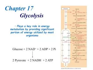
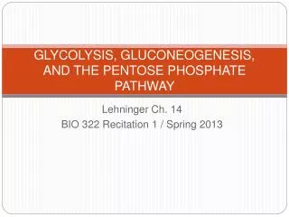
B. Catabolic Pathways • Catabolic reactions serve to capture chemical energy in the form of adenosine triphosphate(ATP) from the degradation of energy-rich fuel molecules. Catabolism also allows molecules in the diet (or nutrient molecules stored in cells) to be converted into building blocks needed for the synthesis of complex molecules. • Energy generation by degradation of complex molecules occurs in three stages
Three stages of metabolism In the first stage, complex molecules are broken down into their component building blocks. For example, proteins are degraded to amino acids, polysaccharides to monosaccharides, and triacylglycerols to free fatty acids and glycerol. In the second stage, these diverse building blocks are further degraded to acetyl coenzyme A (CoA) and a few other, simple molecules. Some energy is captured as ATP, but the amount is small compared with the energy produced during the third stage of catabolism. The tricarboxylic acid (TCA) cycle is the final common pathway in the oxidation of fuel molecules such as acetyl CoA. Large amounts of ATP are generated as electrons flow from NADH and FADH2 to oxygen via oxidative phosphorylation
C. Anabolic pathways • Anabolic reactions combine small molecules, such as amino acids, to form complex molecules, such as proteins (Figure 8.4). • Anabolic reactions require energy (are endergonic), which is generally provided by the breakdown ofATP to adenosine diphosphate (ADP) and inorganic phosphate (Pi). • Anabolic reactions often involve chemical reductions in which the reducing power is most frequently provided by the electron donor NADPH. • catabolism is a convergent process—that is, a wide variety of molecules are transformed into a few common end products. • By contrast, anabolism is a divergent process in which a few biosynthetic precursors form a wide variety of polymeric or complex products.
II. Regulation of Metabolism • The pathways of metabolism must be coordinated so that the production of ENERGY or the synthesis of end products meets the needs of the cell. • Furthermore, individual cells do not function in isolation but, rather are part of a community of interacting tissues. • Thus, a sophisticated COMMUNICATION SYSTEM has evolved to coordinate the functions of the body. • REGULATORY SIGNALS that inform an individual cell of the metabolic state of the body as a whole include hormones, neurotransmitters, and the availability of nutrients. These, in turn, influence signals generated within the cell (Figure 8.5).
A. Signals from within the cell (intracellular) • The rate of a metabolic pathway can respond to regulatory signals that arise from within the cell. • For example, the rate of a pathway may be influenced by the availability of substrates,product inhibition, or alterations in the levels of allosteric activators or inhibitors. • These intracellular signals typically elicit rapid responses, and are important for the moment-to-moment regulation of metabolism.
B. Communication between cells (intercellular) • The ability to respond to extracellular signals is essential for the survival and development of all organisms. • Signaling between cells provides for long-range integration of metabolism, and usually results in a response that is slower than is seen with signals that originate within the cell. • Communication between cells can be mediated by surface-to-surface contact and, in some tissues, by formation of gap junctions, allowing direct communication between the cytoplasms of adjacent cells. • However, for energy metabolism, the most IMPORTANT ROUTE of communication is chemical signaling between cells, for example, by blood borne hormones or by neurotransmitters.
Figure 8.5 Some commonly used mechanisms for transmission of regulatory signals between cells.
C. Second messenger systems • Hormones or neurotransmitters can be thought of as signals, and a receptor as a signal detector. • Each component serves as a link in the communication between extracellular events and chemical changes within the cell. • Many receptors signal their recognition of a bound ligand by initiating a series of reactions that ultimately result in a specific intracellular response. • “SECOND MESSENGER” molecules—so named because they intervene between the original messenger (the neurotransmitter or hormone) and the ultimate effect on the cell—are part of the cascade of events that translates hormone or neurotransmitter binding into a cellular response. • Two of the most widely recognized second messenger systems are the calcium/phosphatidylinositol system (see p. 205), and the adenylylcyclase system, which is particularly important in regulating the pathways of intermediary metabolism.
D. Adenylyl cyclase • The recognition of a chemical signal by some membrane receptors, such as the β- and α2-adrenergic receptors,1triggers either an increase or a decrease in the activity of adenylylcyclase (adenylatecyclase). • This is a membrane-bound enzyme that converts ATP to 3′,5′-adenosine monophosphate (also called cyclic AMP or cAMP). • The chemical signals are most often hormones or neurotransmitters, each of which binds to a unique type of membrane receptor. Therefore, tissues that respond to more than one chemical signal must have several different receptors, each of which can be linked to adenylylcyclase. • Certain toxins, such as one produced by Vibriocholerae, can also activate the adenylylcyclase cascade, with potentially disastrous consequences.2 Other toxins, such as one produced by Bordetellapertussis, inhibit the enzyme. • These receptors are characterized by an extracellular ligand-binding region, seven transmembrane helices, and an intracellular domain that interacts with G Proteins (Figure 8.6).
1 - GTP-dependent regulatory proteins: • The effect of the activated, occupied receptor on second messenger formation is not direct but, rather, is mediated by specialized trimeric proteins of the cell membrane. These proteins, referred to as G proteins because they bindguanosine nucleotides (GTP and guanosine diphosphate [GDP]), form a link in the chain of communication between the receptor and adenylyl cyclase. • The inactive form of a G protein binds to GDP (Figure 8.7). The activated receptor interacts with G proteins, triggering an exchange in which GTP replaces GDP. The trimeric G protein then dissociates into an α subunit and a βγ dimer. • The GTP-bound form of the α subunit moves from the receptor to adenylyl cyclase, which is thereby activated. Many molecules of active G protein are formed by one activated receptor. • [Note: The ability of a hormone or neurotransmitter to stimulate or inhibit adenylyl cyclase depends on the type of G protein that is linked to the receptor. One family of G proteins, designated Gs, is specific for stimulation of adenylyl cyclase; another family, designated Gi, causes inhibition of the enzyme (not shown in Figure 8.7).] The actions of the G protein–GTP complex are short-lived because the G protein has an inherent GTPase activity, resulting in the rapid hydrolysis of GTP to GDP. This causes inactivation of the G protein.
Actions of cAMP 2 - Protein kinases: • The next key link in the cAMP second messenger system is the activation by cAMP of a family of enzymes called cAMP-dependent protein kinases, for example, protein kinase A (Figure 8.8). • Cyclic AMP activates protein kinase A by binding to its two regulatory subunits, causing the release of active catalytic subunits. The active subunits catalyze the transfer of phosphate from ATP to specific serine or threonine residues of protein substrates. • The phosphorylated proteins may act directly on the cell's ion channels, or may become activated or inhibited enzymes. • Protein kinase A can also phosphorylate specific proteins that bind topromoter regions of DNA, causing increased expression of specific genes. [Note: Not all protein kinases respond to cAMP; several types of protein kinases are not cAMP-dependent, for example, protein kinase C described on p. 205.] 3 - Dephosphorylation of proteins: The phosphate groups added to proteins by protein kinases are removed by protein phosphatases—enzymes that hydrolytically cleave phosphate esters. This ensures that changes in enzymic activity induced by protein phosphorylation are not permanent. 4 - Hydrolysis of cAMP:cAMP is rapidly hydrolyzed to 5′-AMP by cAMP phosphodiesterase, one of a family of enzymes that cleave the cyclic 3′,5′-phosphodiester bond. 5′-AMP is not an intracellular signalingmolecule. Thus, the effects of neurotransmitter- or hormone-mediated increases of cAMP are rapidly terminated if the extracellular signal is removed. [Note: Phosphodiesterase is inhibited by methylxanthine derivatives, such as theophylline and caffeine]