Download
1 / 1
10 likes | 139 Vues
Frontier Microfocusing Macromolecular Crystallography Beamline (FMX).

E N D
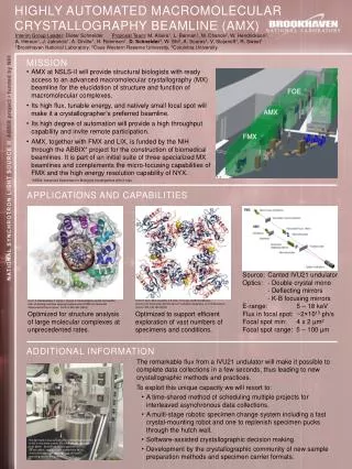
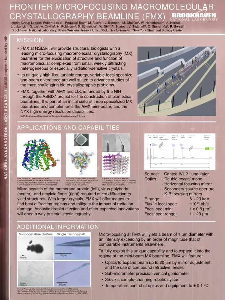
Frontier Microfocusing Macromolecular Crystallography Beamline (FMX) Interim Group Leader: Robert Sweet Proposal Team: M. Allaire1, L. Berman1, M. Chance2, W. Hendrickson3, A. Héroux1, J. Jakoncic1, Q. Liu4, A. Orville1, H. Robinson1, D. Schneider1, W. Shi1, A. Soares1, V. Stojanoff1, D. Stoner-Ma1, R. Sweet1 1Brookhaven National Laboratory, 2Case Western Reserve Univ., 3Columbia University, 4New York Structural Biology Center Mission • FMX at NSLS-II will provide structural biologists with a leading micro-focusing macromolecular crystallography (MX) beamline for the elucidation of structure and function of macromolecular complexes from small, weekly diffracting heterogeneous or especially radiation-sensitive crystals. • Its uniquely high flux, tunable energy, variable focal spot size and beam divergence are well suited to advance studies of the most challenging bio-crystallographic problems. • FMX, together with AMX and LIX, is funded by the NIH through the ABBIX* project for the construction of biomedical beamlines. It is part of an initial suite of three specialized MX beamlines and complements the AMX mini-beam, and the NYX high energy resolution capabilities. • *ABBIX: Advanced Beamlines for Biological Investigations with X-rays APPLICATIONS and Capabilities • Source: Canted IVU21 undulator • Optics: - Double crystal mono • - Horizontal focusing mirror • - Secondary source aperture • - K-B focusing mirrors • E-range: 5 – 23 keV • Flux in focal spot: ~1013 ph/s • Focal spot min: 1 x 0.8 µm2 • Focal spot range: 1 – 20 µm From: T. Warne, M.J. Serrano-Vega, J.G. Baker, R. Moukhametzianov, P.C.Edwards, R. Henderson, A,G. Leslie, C.G. Tate, and G.F. Schertler. Structure of a ß1-adrenergic G-protein coupled receptor. Nature 454, 486-491 (2008). F. Coulibaly, E. Chiu, K. Ikeda, S. Gutmann, P.W. Haebel, C. Schulze-Briese. The molecular organization of cypoviruspolyhedra. Nature 446, 97-101 (2007). R. Nelson, M.R. Sawaya, M. Balbirnie, A.O. Madsen, C. Riekel, R. Grothe, D. Eisenberg. Structure of the cross-beta spine of amyloid-like fibrils. Nature 435, 773-8 (2005). • Micro crystals of the membrane protein (left), virus polyhedra (center) and amyloid fibrils (right) required micro diffraction to • yield structures. With larger crystals, FMX will offer means to • find best diffracting regions and mitigate the impact of radiation damage. Acoustic droplet ejection and other expected innovations will open a way to serial crystallography. Additional Information • Micro-focusing at FMX will yield a beam of 1 µm diameter with an intensity exceeding by an order of magnitude that of comparable instruments elsewhere. • To fully exploit this unique capability and to expand it into the regime of the mini-beam MX beamline, FMX will feature: • Optics to expand beam up to 20 µm by mirror adjustment • and the use of compound refractive lenses • Sub-micrometer precision vertical goniometer • Six-axis sample-changing robotic system • Temperature control of optics and equipment to ± 0.1 ºC From: M.R. Sawaya, S. Sambashivan, R. Nelson, M.I. Ivanova, S.A. Sievers, M.I. Apostol, M.J. Thompson, M. Balbirnie, J.J. Wiltzius, H.T. McFarlane, A.O. Madsen, C. Riekel, and D. Eisenberg. Atomic Structures of Amyloid Cross-ß Spines Reveal Varied Steric Zippers. Nature 447, 453-7 (2007).