Download
1 / 2
20 likes | 123 Vues
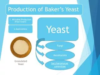
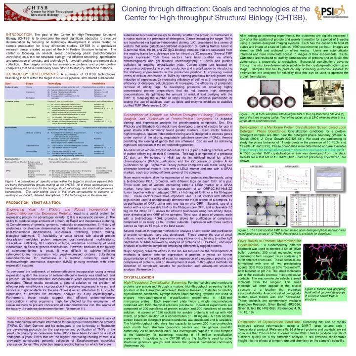
0.5% C 8 E 4 ; 20 C. 0.05% C 12 M; 20 C. CaCl 2. LiCl. 0.4mm. Cloning through diffraction: Goals and technologies at the Center for High-throughput Structural Biology (CHTSB).

E N D
0.5% C8E4; 20 C 0.05% C12M; 20 C CaCl2 LiCl 0.4mm Cloning through diffraction: Goals and technologies at the Center for High-throughput Structural Biology (CHTSB). INTRODUCTION: The goal of the Center for High-Throughput Structural Biology (CHTSB) is to overcome the most significant obstacles to structure determination by focusing on technology development in areas related to sample preparation for X-ray diffraction studies. CHTSB is a specialized research center created as part of the NIH Protein Structure Initiative. The center is focusing on several areas; developing yeast (Saccharomyces cerevisiae) as a tool for structural biology, the efficient screening, optimization and production of crystals, and technology for crystal handling and remote data collection. The targets include transmembrane proteins and protein-protein complexes that have traditionally been difficult to study by diffraction methods. established biochemical assays to identify whether the protein is maintained in a native state in the presence of detergents. Genes encoding the target TMPs are transferred via ligation-independent cloning (LIC) procedures to a series of vectors that allow galactose-controlled expression of reading frames fused to C-terminal His6, His10, and ZZ (IgG-binding) domains that are separated from the reading frame by a cleavage site for rhinovirus 3C protease. Several TMP targets expressed from these vectors have been purified via affinity chromatography and gel filtration chromatography at levels and purities sufficient for ongoing crystallization trials. Current efforts are focused on overcoming bottlenecks in protein production and crystallization by introducing the following improvements in the production pipeline: 1) improving overall levels of cellular expression of TMPs by altering protocols for cell growth and induction of expression; 2) increasing efficiency of cell lysis; 3) increasing the efficiency of detergent solubilization; 4) increasing the efficiency of proteolytic removal of affinity tags; 5) developing protocols for obtaining highly concentrated protein preparations that do not contain high detergent concentrations; 6) optimizing the amount of residual lipid purifying with the TMP; 7) reducing the number of steps required for effective purification; 8) testing the use of additives such as lipids and enzyme inhibitors to stabilize purified TMP (References 6, 31). After setting up screening experiments, the outcomes are digitally recorded 1 day after the addition of protein and weekly thereafter for a period of 6 weeks using 3 custom-designed imaging systems. Each has the capacity to hold 28 plates and image at a rate of 3 plates (4500 experiments) per hour. Images are stored on SAN and archived on offline media. Users are automatically, informed and have immediate access to images of their experimental results. Outcomes are reviewed to identify combinations of proteins and cocktails that demonstrate a propensity to crystallize. Successful combinations advance through the structure-determination pipeline to the crystal-growth optimization stage. Samples that fail to produce any screening outcomes suitable for optimization are analyzed for solubility data that can be used to optimize the protein formulation. TECHNOLOGY DEVELOPMENTS: A summary of CHTSB technologies describing their fit within the target to structure pipeline, with related publications. Goal Steps Technology Availability Produce protein Cloning Develop yeast as a protein production tool: Published methods available to community (1) Overcome Selenomethionine toxicity Reference 11 Expression (2) Produce Transmembrane Proteins Reference 5, 7, 31 (3) Produce Protein Complexes Reference 3 (4) Produce proteins recalcitrant to E. Coli Reference 6, 28 Purification expression Crystallize protein Develop HT tools for crystallization: Published methods and facilities available Screening (1) HT 1536 cocktail soluble protein screen Reference 30 (2) HT 1536 cocktail membrane protein screen Reference 26 (3) ‘Silver bullet’ additive screens potential for Reference 4, 9, 14, 15, 19 crystallization and functional information (4) Develop biophysical techniques to improve and Reference 1 crystallization and determine causes of failure Optimization (1) HT automated DVR/T optimization Reference 8, (2) Crystallization in capettes - in situ X-raydiffraction In progress (3) Software for chemical space map of screening Reference 20, 21, 22, 23 results, automated image analysis (b) (a) Figure 2. (a) A 1536 well plate with enlargements of four crystallization hits and (b) two of the three imaging tables. Two of the tables are at 23oC while the third is in a temperature-controlled room. Development of Methods for Medium-Throughput Cloning, Expression, Analysis, and Purification of Protein:Protein Complexes:To expedite cloning and expression analysis for protein:protein complexes, Drs. Eric Phizicky and Elizabeth Grayhack have developed a suite of vectors for use in yeast strains with commonly found genetic markers. Each vector features high-throughput, ligation-independent cloning and is designed to express genes under control of the strong, regulatablegalactose promoter (PGAL), thereby permitting the cloning of genes that are otherwise toxic as well as achieving high-level expression of the corresponding proteins. An initial set of vectors express individual ORFs (Open Reading Frames) with a tri-partite affinity tag on their C-terminus. This tag is comprised of a protease 3C site, an HA epitope, a His6 tag for immobilized metal ion affinity chromatography (IMAC) purification, and the ZZ domain of protein A for purification on IgGSepharose. Binary protein complexes are made using two otherwise identical vectors (one with a LEU2 marker and one with a URA3 marker), each expressing different genes of the complex. More recent vectors allow for expression of two proteins simultaneously, using a bi-directional PGAL promoter, with different tags on each ORF of a pair. Three such sets of vectors, containing either a LEU2 marker or a URA3 marker, have been constructed for expression of an ORF-3C-HA-His6-ZZ fusion together with an untagged ORF, a His6-tagged ORF, or a His10-tagged ORF. These vectors have three important uses. First, vectors with different tags can be used to unequivocally demonstrate the existence of a complex, by co-purification of ORFs using only one tag on one ORF. Second, use of a vector with a non-cleavable His6 or His10-tag on one ORF, and a cleavable ZZ tag on the other ORF, allows for efficient purification using two affinity steps, each directed at one ORF of the complex. Third, use of pairs of vectors, each with a bi-directional PGAL promoter, allows for purification of complexes comprised of as many as four protein subunits. Expression with these vectors can be as high as 15 mg/L in the best cases. Several medium-throughput methods for analysis of expression and purification of protein complexes have also developed. These employ the use of small cultures, rapid analysis of expression using stick-and-strip binding to either IgGSepharose or IMAC followed by analysis of proteins on SDS-PAGE, and rapid analysis of authentic complexes employing differentially tagged proteins. Major ongoing research efforts in the lab are focused on the development of methods to further enhance expression of proteins in yeast, on further documentation of the utility of yeast for expression of exogenous proteins and complexes of proteins, and on development of medium-throughput methods for detection of complexes suitable for purification and subsequent structural analysis (Reference 3). Development of a Membrane Protein Crystallization Screen based upon Detergent Phase Boundaries: Crystallization conditions for a protein-detergent complex are often near the detergent phase boundary (Wiener & Snook (2001) J. Cryst Growth 232:426-431). We used dye-partitioning to study the phase behavior of 11 detergents in the presence of 10 PEGs and 11 salts (4oand 23oC). Phase boundaries were determined and are available (https://secure.hwi.buffalo.edu/hwi/faculty_research/crystallization/SlickSpot.html). A 1536 cocktail TMP crystallization screen was developed from this data. Results for a test set of 12 TMPs (10/12 had not previously crystallized) are shown. Structural data Develop tools for crystal-handling, Published methods and facilities available data collection, SAXS data analysis Diffraction screening (1) Automated web-enabled home X-ray prescreen : In progress -rapid feedback to crystallization -make optimum use of synchrotron (2) Automated remote room-temperature Available data collection at home and SSRL (Stanford Synchrotron Radiation Lightsource) Cryopreservation (1) Cryo-conditions for 1536 soluble protein cocktails Reference 17 (2) High-pressure Helium cryo-cooling Reference 2, 10, 12, 16 Structural data (1) Remote data collection at SSRL Reference 13 (2) Automated sample handling SSRL SAXS Reference 29 (Small Angle X-ray Scattering) Beamline4-2 Structural solution Single crystal X-ray diffraction and SAXS Single crystal X-ray diffraction Data processing Using established, published protocols and software Structure refinement Validation Annotation Single crystal SAXS Data processing (1) Beta-testing sample-loading automation developed by and SAXS beamlines Analysis Dr. HiroTsuruta at SSRL beamline 4-2. available at SSRL Envelope reconstruction (2) Developing and beta-testing tools for semi-automated data Validation processing, analysis and envelope reconstruction/validation Figure 1. A breakdown of specific areas within the target to structure pipeline that are being developed by groups making up the CHTSB. All of these technologies are being developed as tools for the biology, structural biology, and structural genomics communities. The color-coding used in this chart corresponds to sections of similarly color-coded , detailed descriptions of the technologies in the main text. PRODUCTION - YEAST AS A TOOL Engineering Yeast for Efficient and Robust Incorporation of Selenomethionine into Expressed Proteins:Yeast is a useful system for expressing protein. Its advantages include: 1) It is a eukaryotic system, 2) The ability to produce large amounts of protein, 3) Rapid and inexpensive culturing, 4) Completeness of genetic, genomic, metabolic characterization, 5) History of usefulness for structure determination, 6) Similarities to mammalian cells in post-translational modifications, sub-cellular trafficking, protein folding, biological pathways, 7) Availability of yeast strains with altered protein degradation, unfolded protein response, post-translational modifications, intracellular trafficking, 8) Existence of large, interactive community of yeast laboratories, 9) Ease of genetic manipulation. However, because of the toxicity of selenomethionine to yeast, it was difficult to achieve efficient selenomethionine incorporation into yeast-expressed proteins. Substituting selenomethionine for methionine is a method commonly used for multiwavelength anomalous dispersion (MAD) phasing methods with other expression systems. To overcome the bottleneck of selenomethionine incorporation using a yeast expression system the source of selenomethionine toxicity was identified, and a protocol for selenomethionine incorporation in yeast-expressed protein was developed. These results constitute a general solution to the problem of effective selenomethionine incorporation into proteins expressed in yeast, and remove a major obstacle for the use of yeast as an alternative to E. coli for expression of proteins for structural analysis by X-ray crystallography. Furthermore, these results suggest that efficient selenomethionine incorporation in other organisms might be effected by the employment of similar methods to prevent conversion of selenomethionine into the source of the toxicity, Se-adenosylselenomethionine(Reference 11). Figure 3. The 1536 cocktail TMP screen based upon detergent phase behavior was tested against a group of 12 TMPs. Phase data is available for download. Silver Bullets to Promote Macromolecular Crystallization : A fundamentally different approach was used to develop a set of ‘silver bullet’ cocktails. 200 small molecules were combined to form reagent mixes containing 3 to 20 different chemicals. These cocktails are formulated with one of two precipitating agents; 30% PEG 3350, or 50% TacsimateTM, both buffered at pH 7.0. The small molecules within the cocktails promote macromolecular contacts. The macromolecule selects a small molecule from the reagent. This small molecule will often appear in the crystal structure at a location that promotes structural stability. A second set of biologically related silver bullets was also developed. These cocktails are commercially available (Hampton Research, Silver Bullets HR2-096; Silver Bullets Bio HR2-088) (References 4, 9, 14, 15, 19) CRYSTALLIZATION High-Throughput Crystallization Screening:Purified, soluble and membrane proteins are processed through a mature, high-throughput screening facility (located at the Hauptman-Woodward Medical Research Institute) to identify crystallization conditions. Syringe-based liquid-handling systems are used to prepare microbatch-under-oil crystallization experiments in 1536-well microassay plates. Each experiment plate holds a single macromolecule combined with 1536 chemical solutions (cocktails). Individual experiments are composed of 200 nanoliters of protein solution and 200 nanoliters of cocktail solution. A screen of 1536 cocktails for soluble proteins is set up with 450 microLof protein solution (at a concentration of ~10 mg/mL). A 1536 cocktail screen based on detergent phase boundaries was developed and is available. The high-throughput laboratory has capacity to screen 200 macromolecules each month from structural genomics centers and the general scientific community. As of December 2009, 944 investigators supplied 11,689 samples to the laboratory for screening, resulting in 18 million crystallization experiments. In addition to the CHTSB efforts this facility is used by other structural genomics groups and serves the general biomedical community (Reference 29). Figure 4. Mellitic acid ‘grappling hook’ with 6 carboxylate groups in a unique bovine trypsin structure Yeast Trans Membrane Protein Production: To address the severe lack of three-dimensional structural information for eukaryotic transmembrane proteins (TMPs), Dr. Mark Dumont and his colleagues at the University of Rochester are developing protocols for the expression and purification of TMPs in the yeast Saccharomyces cerevisiae. Initial efforts have been focused on a set of endogenous yeast TMPs that are the highest expressing reading frames in a previously constructed genomic collection of Saccharomyces cerevisiae expression clones. This collection targets reading frames for which there are Optimization of Crystallization Conditions:Screening hits can be rapidly optimized without reformulation using a DVR/T (drop volume ratio / Temperature) protocol (Reference 8). 96 different proteins and cocktails are set up in less than 2 hours time. In cases where DVR/T fails to produce crystals of sufficient quality for X-ray diffraction analysis, it still provides considerable insight into the effects of temperature and chemistry on the sample’s solubility.
Remote Single Crystal Data Collection:By working closely with crystallographers at the Hauptman-Woodward Institute (HWI), the Structural Molecular Biology (SMB) group at the Stanford Synchrotron Radiation Lightsource(SSRL) is perfecting the technology for remote access data collection. The SSRL SMB group operates seven beam lines (BL1-5, BL7-1, BL9-1, BL9-2, BL11-1, and BL11-3) for macromolecular crystallography experiments and a new undulator station optimized for microcrystal data collection, BL12-2. The users of these beam lines have the option to collect data remotely and, during the 2007 run period, about 70% of experiments were scheduled for remote access thereby saving travel time and expenses. While remaining at their home institutions, remote users conduct experiments by means of advanced software tools enabling network-based systems monitoring and control. Remote users have the capability to mount, center (Reference13), and screen samples as well as to collect, analyze, and backup diffraction data. Automated sample mounting is accomplished with the Stanford Auto-Mounting System (SAM). In this way, users can screen crystals 2-3 times more rapidly with less human error. For rapid crystal ranking and data analysis, the diffraction images collected during screening are automatically analyzed. Software Developments for Crystallization: Software has been developed to enhance the capabilities, and improve the efficiency of the crystallization laboratory. AutoSherlock,for presenting crystallization screening results in chemical space using color-coded outcomes for ease of interpretation of solubility, crystallization and optimization trends with hyperlinked images for closer review (References 22, 23). Figure 5. DVR/T optimization is set up for 96 proteins simultaneously. This figure shows a single protein with 16 different drop volume ratios of protein to cocktail set up at 5 different temperatures. Columns have a constant drop volume ratio, with temperature as the variable; rows have a constant temperature with drop volume ratio as the variable. An 80 condition optimization requires 25 microL of protein. Image Analysis:There are over 100 million images generated by the screening laboratory that remain unclassified. A training set of images to guide software-controlled classifiers was compiled. Crystallization outcomes from 96 macromolecular samples that underwent 1536 cocktail screening (MW range of 10 – 2000 kDa) were manually classified by 8 reviewers (each image classified by 3 reviewers, with an even distribution of reviewers/image). Of the 147,456 images selected for this study 70, 465 were unanimously classified. Images fell into ten main categories. The distribution of precipitates (41.7%) and clear drops (41.2%) was nearly identical; 49 of the 96 samples had 1 or more outcomes classified as crystals; crystals were a rare occurrence appearing in 0.38% of the images (References 20, 21). Machine classification is taking place at OCI using the human-classified ‘truth data’. Feature extraction that will lead to the development of more accurate classifiers is progressing on The World Community Grid. Currently over 42,000 years of computer processing time has been used for the distributed project. Beamline Emulator: An automated set up that provides rapid X-ray based feedback to the crystal optimization has been constructed at HWI. The system mimics the SSRL Stanford Automated Mounting Systems (SAM) (Reference 30). Based on typical data collection times 2 cassettes of samples, 192 in total, will be screened in the laboratory and sorted in ~70 hours. This pre-screening will guide crystal optimization (based on diffraction quality) and maximize productive use of beamtime at SSRL. not sampled precipitate clear crystal Figure 9. SAM system installed at HWI with Mar 345 imaging plate detector. Figure 7. AutoSherlock which is available to the general biomedical community to analyze their crystallization screening results. DATA MINING Databases and Information Systems: Data mining in all areas is under development. Data mining on a subset of crystallization results was completed and published (References 20,21). The aim is to expand the analysis developed on this subset to analyze the full archive of results. X-RAY STUDIES Small Angle X-ray Scattering (SAXS): To complement crystallization studies we have made use of the SAXS method and are working on developing a high-throughput pipeline in collaboration with Hiro Tsuruta at SSRL. Table 1. Performance of current classifier. Precipitated and clear drops on average constitute 83% (1275 out of 1536) outcomes of the HT crystal screen. If we can focus our efforts on the 17% (261 out of 1536) images that are most likely to contain crystals, we can eliminate time-consuming manual review of the less-interesting outcomes. By reordering the images, placing clear and precipitated drops after the more-interesting outcomes, we can significantly improve our efficiency and throughput. By adding a time component we further improve classification. Examples of envelopes where we have known structures and where SAXS also reveals residues that were not resolved in the crystal structure Correcting crystallographic packing artifacts Figure 10. Early data mining results from scored crystallization trials. REFERENCES 1. Ericsson, U. B., Hallberg, B. M., Detitta, G. T., Dekker, N., and Nordlund, P. (2006). Thermofluor-based high-throughput stability optimization of proteins for structural studies. Analytical Biochemistry, 357: 289-298. [PubMed ID: 16962548] 2. Kim, C., Hao, Q., and Gruner, S.M. (2006). Solution of Protein Crystallographic Structures by High-Pressure Cryocooling and Noble-Gas Phasing. ActaCryst.D62: 687-694. [Pub Med ID: 16790924] 3. Phizicky, E. M. and Grayhack, E. J. (2006). Proteome-Scale Analysis of Biochemical Activity. Crit. Rev. Biochem. Mol. Biol.41: 315-327. [Pub Med ID: 16911958] 4. McPherson, A. and Cudney, R. (2006). Searching for Silver Bullets: An Alternative Strategy for Crystallizing Macromolecules. J. Structural Biology156: 387-406. [Pub Med ID: 17101277] 5. Wang, Z., Stalcup, L. D., Harvey, B. J, Weber, J., Chloupkova, M., Dumont, M. E., Dean, M., and Urbatsch, I. L. (2006). Purification and ATP hydrolysis of the putative cholesterol transporters ABCG5 and ABCG8. Biochemistry, 45: 9929-9939. [Pub Med ID: 16893193] 6. White, M. A., Clark, K. M., Grayhack, E. J., and Dumont, M. E. (2007). Characteristics Affecting Expression and Solubilization of Yeast Membrane Proteins. J. Mol. Biol.365: 621-636. [Pub Med ID:17078969] 7. Chloupková, M., Pickert, A., Lee, J. Y., Souza, S., Trinh, Y. T, Connelly, S. M., Dumont, M. E., Dean, M,, and Urbatsch, I. L. (2007). Expression of 25 human ABC transporters in the yeast Pichiapastoris and characterization of the purified ABCC3 ATPase activity. Biochemistry, 46: 7992-8003. [Pub Med ID: 17569508] 8. Luft, J. R., Wolfley, J. R., Said, M. I., Nagel, R. M., Lauricella, A. M., Smith, J. L., Thayer, M. H., Veatch, C. K., Snell, E. H., Malkowski, M. G., and DeTitta, G. T. (2007). Efficient Optimization of Crystallization Conditions by Manipulation of Drop Volume Ratio and Temperature. Protein Science,16: 715-722. [Pub Med ID: 17327388] 9. Larson, S. B., Day, J. S., Cudney, R., and McPherson, A. (2007). A Novel Strategy for the Crystallization of Proteins: X-ray Diffraction Validation. ActaCryst. D63: 310-318. [Pub Med ID:17327668] 10. Collins, M.C., Quillin, M. L., Hummer, G., Matthews, B. W., and Gruner, S. M. (2007). Structural Rigidity of a Large Cavity-Containing Protein Revealed by High-Pressure Crystallography. J. Mol. Biol.367: 752-763. [Pub Med ID: 17292912] 11. Malkowski, M. G., Quartley, E., Friedman, A. E., Babulski, J., Kon, Y., Wolfley, J., Said, M., Luft, J. R., Phizicky, E. M., DeTitta, G. T., and Grayhack, E. J. (2007). Blocking S-Adenosylmethionine Synthesis in Yeast Allows Selenomethionine Incorporation and Multiwavelength Anomalous Dispersion Phasing. Proc. Natl. Acad. Sci., USA.104: 6678-6683. [Pub Med ID: 17426150] 12. Kim, C., Hao, Q., and Gruner, S. M. (2007). High Pressure Cryocooling for Capillary Sample Cryoprotection and Diffraction Phasing at Long Wavelengths. ActaCryst.D63: 653-659. [Pub Med ID:17452791] 13. Song, J., Mathew, D., Jacob, S. A., Corbett, L., Moorhead, P., and Soltis, S. M.. (2007). Diffraction-based Automated Crystal Centering. J. Synchrotron Rad.14: 191-195. [Pub Med ID: 17317920] 14. Larson, S. B., Day, J. S., Cudney, R., and McPherson, A. (2007). A New Crystal Form of Bovine Pancreatic RNase A in Complex with 2'-Deoxyguanosine-5'-monophosphate. ActaCrystallogr. Sect. F Struct. Biol. Cryst. Commun.63: 728-733. [Pub Med ID: 17768339] 15. McPherson, A., Nguyen, C., Larson, S.B., Day, J.S., and Cudney, B. (2007). Development of an Alternative Approach to Protein Crystallization. J. Struct. Funct. Genomics.8: 193-198. [Pub Med ID:18038192] 16. Kim, C. U., Chen, Y.-F., Tate, M. W., and Gruner, S. M. (2008). Pressure Induced High-density Amorphous Ice in Protein Crystals. J. Appl. Cryst.41: 1-7. 17. Kempkes, R., Stofko, E., Lam, K., and Snell, E. H. (2008). Glycerol concentrations required for the successful vitrification of cocktail conditions in a high-throughput crystallization screen. ActaCrystallogr D BiolCrystallogr.64: 287-301. [Pub Med ID: 18323624] 18. Fox, B. G., Goulding, C., Malkowski, M. G., Stewart, L., and Deacon, A. (2008). Structural genomics: from genes to structures with valuable materials and many questions in between. Nature Methods,5: 129-132. [Pub Med ID: 18235432] 19. Larson, S.B., Day, J.S., Nguyen, C., Cudney, R., and McPherson, A. (2008) Progress in the development of an alternative approach to macromolecular crystallization. Crystal Growth and Design,8: 3038-3052. [URL] 20. Snell, E. H., Luft, J. R., Potter, S. A., Lauricella, A. M., Gulde, S. M., Malkowski, M. G., Koszelak-Rosenblum, M., Said, M. I., Smith, J. L., Veatch, C. K., Collins, R. J., Franks, G., Thayer, M., Cumbaa, C., Jurisica, I., and DeTitta, G. T. (2008). Establishing a training set through the visual analysis of crystallization trials part I: ~150,000 images. ActaCryst. D64: 1121-1130. [Pub Med ID: 19020350] 21. Snell, E. H., Lauricella, A. M., Potter, S. A., Luft, J. R., Gulde, S. M., Collins, R. J., Franks, G., Malkowski, M. G., Cumbaa, C., Jurisica, I., and DeTitta, G. T. (2008). Establishing a training set through the visual analysis of crystallization trials part II: Crystal examples. ActaCryst. D64: 1131-1137. [Pub Med ID: 19020351] 22. Nagel, R. M., Luft, J. R., and Snell, E. H. (2008). AutoSherlock: a program for effective crystallization data analysis. J. Appl. Cryst. 41: 1173-1176. [URL] 23. Snell, E. H., Nagel, R. M., Wojtaszcyk, A., O'Neill, H., Wolfley, J., and Luft, J. R. (2008). The application and use of Chemical Space Mapping to interpret crystallization screening results. ActaCryst. D64: 1240-1249. [Pub Med ID: 19018100] 24. Price, W. N. 2nd, Chen, Y., Handelman, S. K., Neely, H., Manor, P., Karlin, R., Nair, R., Liu, J., Baran, M., Everett, J., Tong, S. N., Forouhar, F., Swaminathan, S. S., Acton, T., Xiao, R., Luft, J. R., Lauricella, A., Detitta, G. T., Rost, B., Montelione, G. T., and Hunt, J. F. (2008). Understanding the physical properties that control protein crystallization by analysis of large-scale experimental data. Nature Biotechnology, 27: 51-57. [Pub Med ID: 19079241] 25. Larson, S. B., Day, J. S., Nguyen, C., Cudney, R., and McPherson, A. (2009). High-resolution structure of proteinase K cocrystallized with digalacturonic acid. ActaCryst.F65: 192-198. [Pub Med ID: 19255463] 26. Koszelak-Rosenblum, M., Krol, A., Mozumdar, N., Wunsch, K., Ferin, A., Cook, E., Veatch, C. K., Nagel, R., Luft, J. R., Detitta, G. T., and Malkowski, M. G. (2009). Determination and application of empirically derived detergent phase boundaries to effectively crystallize membrane proteins. Protein Sci.18: 1828-1839. [Pub Med ID: 19554626] 27. Larson, S. B., Day, J. S., Nguyen, C., Cudney, R., and McPherson, A. (2009). Structure of pig heart citrate synthase at 1.78Å resolution. ActaCrystallogr. Sect. F Struct. Biol. Cryst. Commun.65: 430-434. [Pub Med ID: 19407370] 28. Quartley, E., Alexandrov, A., Mikucki, M., Buckner, F. S., Hol, W. G., DeTitta, G. T., Phizicky, E. M., and Grayhack, E. J. (2009). Heterologous expression of L. major proteins in S. cerevisiae: a test of solubility, purity, and gene recoding. J. Struct. Funct. Genomics. 10: 233-247. [Pub Med ID: 19701618] 29. Luft et al., (2003).A deliberate approach to screening for initial crystallization conditions of biological macromolecules. J. Struct. Biol. 142, 170-179. 30. Cohen et al., (2002). An automated system to mount cryo-cooled protein crystals on a synchrotron beamline, using compact sample cassettes and a small scale robot. J. Appl. Cryst. 35, 720-726. 31. Clark, K.M., Fedoriw, N., Robinson K, Connelly, S.M., Randles, J., Malkowski, M.G., DeTitta, G.T., Dumont, M.E., (2010) Purification of Transmembrane Proteins from Saccharomyces cerevisiae for X-ray Crystallography. Protein Expression and Purification. Accepted for Publication Crystal Production, Cryopreservation, and Diffraction in Capillaries: We are developing pipette tips as vessels for crystal growth cryopreservation, and in situ X-ray diffraction. This will improve efficiency and eliminate potential damage caused by physical manipulation. This work is a collaboration between SSRL, HWI, and Cornell. Our collaborators at SSRL designed a pipette tip for compatibility with HWI liquid-handling systems and the automated sample handling system at SSRL. Tips produced from a number of different plastics were tested for X-ray diffraction properties (SSRL), compatibility with robotics (HWI, SSRL) (Reference 30), crystallization (HWI), and cryo-compatibility (Cornell). Large scale production of tips is now underway. High through-put studies and resulting ‘unfiltered’ quality of the data. Automatic routines are being developed to flag cases that need expert intervention (seen as high chi values and ‘exploded’ envelopes). An example of one of our targets, Gln4, where 214 residues were missing in the crystallographic structure but resolved in the SAXS work and confirmed by low resolution crystallographic analysis (the system has an ordered but highly flexible arm) Figure 8. Examples of our SAXS studies showing the high-throughput envelope reconstruction (without filtering techniques applied) and some of the applications of the current pipeline complementing crystallographic approaches. SUMMARY: CHTSB has been focused on technology developments. These developments are now bearing fruit in terms of production, identification of crystallization leads, optimization and diffraction. The tools developed feed into the corresponding pipeline from target to structure but individually are applicable and available to the wider general biomedical community. Figure 6. Capette development showing (a) a schematic of the plastic capillary/tip, (b) the tip positioned within a uni-puck illustrating how the form factor is compatible with many systems that utilize a standard magnetic cap, (c) an early test example with a crystal within the tip (and an enlarged image of the crystal) (d) diffraction from a crystal within one of the plastic tips cryocooled under pressure (see later section). (a) (b) ACKNOWLEDGEMENTS: Work supported by NIH U54 GM074899, the John R. Oishei Foundation, Margaret L. Wendt Foundation, Erie County and the James H. Cummings Foundation. People (c) (d) Yoshiko Kon Maryann Mikucki Eric Phizicky Erin Quartley Katrina Robinson Kathy Clark Sara Connelly Mark Dumont Nadia Fedoriw Elizabeth Grayhack Eleanor Cook George DeTitta Christopher Goulah Thomas Grant Angela Lauricella Joseph Luft Michael Malkowski Raymond Nagel Walter Pangborn Stephen Potter Mary Rosenblum Edward Snell Elizabeth Snell Max Thayer Christina Veatch Charles Weeks Jennifer Wolfley Department of Molecular Biology and Biochemistry, University of California, 560 SH, Mail Code 3900 Irvine, CA 92697 Building 120 SLAC 2575 Sand Hill Road Menlo Park, CA 94025 Christian Cumbaa Igor Jurisica Department of Physics/CHESS, 162 Clark Hall, Cornell University, Ithaca NY 14853 Division of Signaling Biology, Ontario Cancer Institute, Princess Margaret Hospital, 610 University Avenue, Toronto, ON, M5G 2M9 John Day Aaron Greenwood Steven Larson YriKuznetsov Alexander McPherson Yi-Fan Chen Sol Gruner Chae Un Kim TzankoDoukov Keith Hodgson Michael Soltis Joseph Chang Aina Cohen