Download
1 / 21
300 likes | 943 Vues
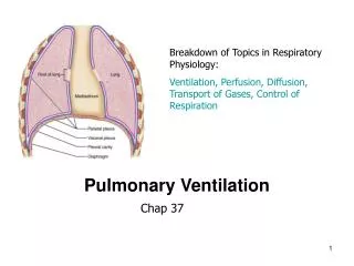
Respiratory Physiology: Control of ventilation. Lecture 15 Tuesday, February 13, 2007 Refs. Medical Physiology Chapters 30 and 31, Ganong, Tadlock. Control of ventilation. Automatic rhythm of ventilation is controlled by a central pattern generator (CPG) in the brain stem.

E N D
Respiratory Physiology:Control of ventilation Lecture 15 Tuesday, February 13, 2007 Refs. Medical Physiology Chapters 30 and 31, Ganong, Tadlock
Control of ventilation • Automatic rhythm of ventilation is controlled by a central pattern generator (CPG) in the brain stem. • In general, CPGs have a set of cyclic, coordinated timing signals that are generated by a cluster of interconnected neurons. These signals command many muscles to contract or relax at a specific phase in the cycle (e.g. walking, running, etc.). • The specific site for the generation of the automatic respiratory rhythm is still unknown. • Adjustments in the basic rhythm are made for changing metabolic demands, changes in body position and talking, eating, etc.
Methods to study control of ventilation • Surgical transections and lesions • Electrical stimulation of brain areas • Recording electrical activity • Early work with surgical lesions was not repeatable and electrical stimuli were very large probably stimulating regions not individual cells. • Through electrical recording, a large number of respiratory-related neurons (RRN) have been identified in many areas of the brain. • Interneurons • Premotor neurons • Motor neurons
Functional mapping of RRNs • Functional mapping has produced a more detailed map of areas involved in the control of respiration. • The dorsal respiratory group DRG sends motor neurons to the primary muscles of inspiration (diaphragm and external intercostal muscles). • The ventral respiratory group VRG previously thought to be involved in active expiration has areas involved with normal inspiration and accessory inspiration and expiration (Table 31-2).
Possible role of pre-Bötzinger complex as the respiratory central-pattern generator MP 31-8
Chemoreceptors for respiration • Peripheral receptors are sensitive to an increase in PCO2 and [H+] or decrease in arterial PO2 • carotid bodies • aortic bodies • Central receptors are sensitive to increased PCO2 and [H+] in blood, CSF, and brain interstitial fluid • ventral surface of the medulla • additional locations include solitary tract, locus ceruleus, and hypothalamus
Location of peripheral chemoreceptors (carotid and aortic bodies)Ganong 36-4
Type I and II cells of the carotid body Ganong 36-5 When exposed to hypoxia, Glomus type I cells secrete catecholamines that stimulate the endings of glossopharyngeal afferent axons. Impulses ascend to the medulla and increase ventilation.
Increased respiration in subjects breathing oxygen and 2, 4, or 6% CO2Ganong 36-8 Demonstrates that rate and depth of respiration is more sensitive to PCO2 than PO2.
Chemoreceptors for respiration • Peripheral receptors are sensitive to an increase in PCO2 and [H+] or decrease in arterial PO2 • carotid bodies • aortic bodies • Central receptors are sensitive to increased PCO2 and [H+] in blood, CSF, and brain interstitial fluid • ventral surface of the medulla • additional locations include solitary tract, locus ceruleus, and hypothalamus
Metabolic functions of the lung • Synthesis of surfactant • Synthesis or release of mediators • prostaglandins, histamine, and kallikrein • Inactivation/removal of mediators • prostaglandins, serotonin, bradykinin, ACh, norepinephrine,adenine nucleotides from blood • Activation of precursor • angiotensin I converted to angiotensin II