Download

1 / 63
630 likes | 866 Vues
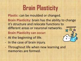
Chapter 5 Development and Plasticity of the Brain. Development of the Brain. Plasticity of the brain refers the the idea that the brain is constantly changing throughout the lifetime. Development of the brain is due to both experience and physical maturation.

E N D
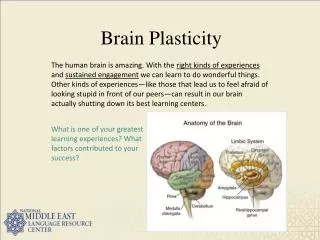
Development of the Brain • Plasticity of the brain refers the the idea that the brain is constantly changing throughout the lifetime. • Development of the brain is due to both experience and physical maturation. • Rapid development especially occurs early in life. • Prefrontal cortex develops rapidly between 7 and 12 months (allowing for object permanence).
Development of the Brain. • The human central nervous system begins to form when the embryo is approximately 2 weeks old. • The dorsal surface thickens forming a neural tube surrounding a fluid filled cavity. • The forward end enlarges and differentiates into the hindbrain, midbrain and forebrain. • The rest of the neural tube becomes the spinal cord.
Development of the Brain • The fluid-filled cavity becomes the central canal of the spinal cord and the four ventricles of the brain. • The fluid is the cerebrospinal fluid.
Development of the Brain • At birth, the human brain weighs approximately 350 grams. • By the first year. the brain weighs approximately 1000 grams. • The adult brain weighs 1200-1400 grams.
Development of the Brain • The development of neurons in the brain involves the following four processes: • Proliferation • Differentiation • Myelination • synaptogenesis
Development of the Brain • Proliferation refers to the production of new cells/ neurons in the brain primarily occurring early in life. • Early in development, the cells lining the ventricles divide. • Some cells become stem cells that continue to divide • Others remain where they are or become neurons or glia that migrate to other locations.
Development of the Brain • Migration refers to the movement of the newly formed neurons and glia to their eventual locations. • Migration occurs in a variety of directions throughout the brain. • Migration occurs via cells following chemical paths in the brain of immunoglobins and chemokines.
Development of the Brain • Differentiation refers to the forming of the axon and dendrite that gives the neuron its distinctive shape. • The axon grows first either during migration or once it has reached its target and is followed by the development of the dendrites. • Neurons differ in their shape and chemical component depending on their location in the brain.
Development of the Brain • Myelination refers to process by which glia produce the fatty sheath that covers the axons of some neurons. • Myelin speeds up the transmission of neural impulses. • Myelination first occurs in the spinal cord and then in the hindbrain, midbrain and forebrain. • Myelination occurs gradually for decades.
Development of the Brain • Synaptogenesis is the final stage of neural development and refers to the formation of the synapses between neurons. • Occurs throughout the life as neurons are constantly forming new connections and discarding old ones. • Synaptogenesis slows significantly later in the lifetime.
Development of the Brain • Although it was originally believed that no new neurons were formed after early development, recent research suggests otherwise: • Two Examples: • Stem cells are undifferentiated cells found in the interior of the brain that generate “daughter cells” which can transform into glia or neurons. • New olfactory receptors also continually replace dying ones.
Development of the Brain • Animal research has also shown the development of new neurons occurring in other brain regions. • Example: songbirds have a steady replacement of new neurons in the singing area of the brain.
Development of the Brain • Axons must travel great distances across the brain to form the correct connections. • Sperry’s (1954) research with newts indicated that axons follow a chemical trial to reach their appropriate target. • Growing axons reach their target area by following a gradient of chemicals in which they are attracted by some chemicals and repelled by others.
Development of the Brain • When axons initially reach their targets, they form synapses with several cells. • Postsynaptic cells strengthen connection with some cells and eliminate connections with others. • The formation or elimination of these connections depends upon input from incoming of axons.
Development of the Brain • Some theorists refer to the idea of the selection process of neural connections as neural Darwinism. • In this competition amongst synaptic connections, we initially form more connections than we need. • The most successful axon connections and combinations survive while the others fail to sustain active synapses.
Development of the Brain • A neurotropin is a chemical that promotes the survival and activity of neurons. • Axons that are not exposed to neurotropins after making connections undergo apoptosis, a preprogrammed mechanism of cell death. • Therefore, the healthy adult nervous system contains no neurons that failed to make appropriate connections.
Development of the Brain • Nerve growth factor (NGF) is a type of neurotrophin released by muscles that promotes the survival and growth of axons. • The brain’s system of overproducing neurons and then applying apoptosis enables the exact matching of the number of incoming axons to the number of receiving cells.
Development of the Brain • The elimination and period of massive cell death is part of normal development and maturation. • After maturity, the apoptotic mechanisms become dormant. • Neurons no longer need neurotrophins for survivals, but neurotrophins increase the branching on axons and dendrites throughout life.
Development of the Brain • Early stages of brain development are critical for normal development later in life. • Chemical distortions in the brain during early development can cause significant impairment and developmental problems.
Development of the Brain • Fetal alcohol syndrome is a condition that children are born with if the mother drinks heavily during pregnancy. • The condition is marked by the following: • Hyperactivity and impulsiveness • difficulty maintaining attention • varying degrees of mental retardation • motor problems and heart defects • facial abnormalities.
Development of the Brain • The dendrites of children born with fetal alcohol syndrome are short with few branches. • Exposure to alcohol in the fetus brain suppresses glutamate and enhances the release of GABA. • Many neurons consequently receive less excitation and exposure to neurotrophins than usual and undergo apoptosis.
Development of the Brain • Children of mothers who use cocaine during pregnancy show a decrease in language skills, a slight decrease in IQ scores and impaired hearing. • Children of mothers who smoked during pregnancy are at increased risk for low birth weight, sudden infant death syndrome, ADHD, long term intellectual deficits and impairments of the immune system.
Development of the Brain • The brain has the limited ability to reorganize itself in response to experience. • Axons and dendrites continue to modify their structure and connections throughout the lifetime. • Dendrites continually grow new spines. • The gain and loss of spines indicates new connections and potentially new information processing.
Development of the Brain • Rats raised in an enriched environment develop a thicker cortex and increased dendritic branching. • Measurable expansion of neurons has also been shown in humans as a function of physical activity. • The thickness of the cerebral cortex declines in old age but much less in those that are physically active.
Development of the Brain • Neurons also become more finely tuned and responsive to experiences that have been important in the past. • This may account for the fact that blind people often have enhanced tactile senses and increased verbal skills. • The occipital lobe normally dedicated to processing visual information adapts to also process tactile and verbal information.
Development of the Brain • Extensive practice of a skill changes the brain in a way that improves the ability for that skill. • For example, MRI studies reveal following: • the temporal lobe of professional musicians in the right hemisphere is 30% larger than non-musicians. • thicker gray matter in the part of the brain responsible for hand control and vision of professional keyboard players
Development of the Brain • Part of the mechanism on increased ability due to experience is that attention to anything releases dopamine. • Dopamine acts on the cortex to expand the response to stimuli active during the dopamine release.
Development of the Brain • Focal hand dystonia or “musicians cramp” refers to a condition where the reorganization of the brain goes too far. • The fingers of musicians who practice extensively become clumsy, fatigue easily and make involuntary movements. • This condition is a result of the area of the brain responsible for a specific finger movement growing and overlapping with others.
Plasticity After Brain Damage • Survivors of brain damage show subtle to significant behavioral recovery. • Some of the mechanisms of recovery include those similar to the mechanisms of brain development such as the new branching of axons and dendrites.
Plasticity After Brain Damage • Possible causes of brain damage include: • Tumors • infections • exposure to toxic substances • degenerative diseases • closed head injuries.
Plasticity After Brain Damage • A closed head injury refers to trauma that occurs when a sharp blow to the head drives the brain tissue against the inside wall of the skull. • One of the main causes of brain injury in young adults • A stroke or cerebrovascular accident is temporary loss of blood flow to the brain. • A common cause of brain damage in the elderly
Plasticity After Brain Damage • Types of strokes include: • Ischemia -the most common type of stroke, resulting from a blood clot or obstruction of an artery. Neurons lose their oxygen and glucose supply. • Hemorrhage -a less frequent type of stroke resulting from a ruptured artery. Neurons are flooded with excess excess calcium oxygen and other products.
Plasticity After Brain Damage • Ischemia and hemorrhage also cause: • Edema-the accumulation of fluid in the brain resulting in increased pressure on the brain and increasing the probability of further strokes. • Disruption of the sodium-potassium pump leading to the accumulation of potassium ions inside neurons.
Plasticity After Brain Damage • Edema and excess potassium triggers the release of the excitatory neurotransmitter glutamate. • The overstimulation of neurons leads to sodium and other ions entering the neuron in excessive amounts. • Excess positve ions in the neuron block metabolism in the mitochondria and kill the neuron.
Plasticity After Brain Damage • A drug called tissue plasminogen activator (tPA) breaks up blood clots and reduces the effects of an ischemic strokes. • Research has begun to attempt to save cells in the penumbra or region that surrounds the immediate damage by: • blocking glutamate synapses • opening potassium channels
Plasticity After Brain Damage • One of the most effective laboratory methods used to minimize damage caused by strokes is to cool the brain. • A cooled brain (91-97° F) has less activity, lower energy needs and less risk of overstimulation.
Plasticity After Brain Damage • Cannabanoids have also been shown to potentially minimize cell loss after brain damage be decreasing the release of glutamate. • Excess glutamate may result in the over-excitation of neurons.