Download
1 / 20
200 likes | 376 Vues
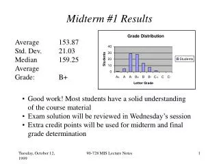
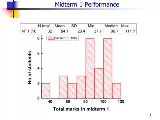
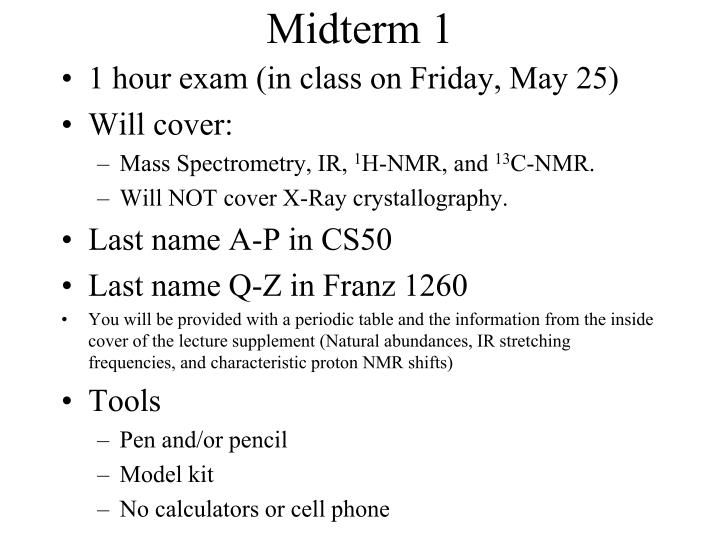
Midterm 1. 1 hour exam (in class on Friday, May 25) Will cover: Mass Spectrometry, IR, 1 H-NMR, and 13 C-NMR. Will NOT cover X-Ray crystallography. Last name A-P in CS50 Last name Q-Z in Franz 1260

E N D
Midterm 1 • 1 hour exam (in class on Friday, May 25) • Will cover: • Mass Spectrometry, IR, 1H-NMR, and 13C-NMR. • Will NOT cover X-Ray crystallography. • Last name A-P in CS50 • Last name Q-Z in Franz 1260 • You will be provided with a periodic table and the information from the inside cover of the lecture supplement (Natural abundances, IR stretching frequencies, and characteristic proton NMR shifts) • Tools • Pen and/or pencil • Model kit • No calculators or cell phone
How should I study? • Review past “Exam 2”s on Hardinger’s websitehttp://www.chem.ucla.edu/harding/index.html(on left frame, click “Ch14C” then in middle frame click “Current and Past Exam and Keys”) Note: X-ray crystallography will not be on Midterm 2, so please skip those questions on the practice exams
13C-NMR, 2D-NMR, and MRILecture Supplement page 181 An NMR spectrometer An MRI instrument
13C-NMR Is NMR limited to 1H? • Any nucleus with I (spin quantum number) 0 can be studied by NMR • I 0 when nucleus has odd number of protons or odd number of neutrons • Includes 1H, 2H, 13C, 19F, 29Si, 31P, 127I, etc. • Examples • 19F: 9 protons, 10 neutrons; 100% natural abundance • 31P: 15 protons, 16 neutrons; 100% natural abundance • 19F, 31P easily observed by NMR, but of limited value for organic structure analysis • 13C-NMR • 13C: 6 protons, 7 neutrons; I 0, 1.1% natural abundance • Carbon is backbone of organic molecules so 13C-NMR very useful
13C-NMR What can we deduce about molecular structure from 13C-NMR spectrum? • NMR fundamentals are the same regardless of nucleus • Information from 13C-NMR spectrum • 1. Number of signals: Equivalent carbons and molecular symmetry • 2. Chemical shift: Presence of high EN atoms or pi electron clouds • 3. Integration: Ratios of equivalent carbons • 4. Coupling: Number of neighbors
13C-NMR: Number of Signals Number of 13C-NMR signals reveals equivalent carbons • One signal per unique carbon type • Reveals molecular symmetry • Examples: CH3CH2CH2CH2OH No equivalent carbons Four 13C-NMR signals CH3CH2OCH2CH3 2 x CH3 equivalent 2 x CH2 equivalent Two 13C-NMR signals If # of 13C-NMR signals < # of carbons in formula then molecule has some symmetry
13C-NMR: Position of Signals • Position of signal relative to reference = chemical shift • 13C-NMR reference = TMS = 0.00 ppm • 13C-NMR chemical shift range = 0 - 250 ppm • Deshielding caused by electronegative atoms and pi electron clouds Example: HOCH2CH2CH2CH3 OH does not have carbon no 13C-NMR signal for OH
13C-NMR: Position of Signals • Trends • EN atoms cause deshielding It is not necessary to memorize this table. It will be given on an exam if necessary. • RCH3 < R2CH2 < R3CH EN C > EN H • Pi bonds cause deshielding and shielding • C=O 160-220 ppm Cross-check with IR zone 4
13C-NMR: Integration 1H-NMR: Integration reveals relative number of hydrogens per signal • 13C-NMR: Integration reveals relative number of carbons per signal • Rarely useful due to slow relaxation time for 13C • Relaxation time: Time for nucleus to relax from excited spin state to ground state I = - ½ Excited state I = + ½ Ground state • 1H relaxation time important phenomenon for MRI
1H 13C 13C 12C 13C-NMR: Spin-Spin Coupling Spin-spin coupling of nuclei causes splitting of NMR signal • Only nuclei with I 0 can couple • Examples: 1H couples with 1H, 1H couples with 13C, 13C couples with 13C • 1H does not couple with 12C • 1H NMR: Splitting reveals number of H neighbors • 13C-NMR: Limited to nuclei separated by just one sigma bond; no pi bond “free spacers” Coupling occurs No coupling: Too far apart Coupling occurs but signal very weak: Low probability for two adjacent 13C 1.1% x 1.1% = 0.012% No coupling: 12C has I = 0 • Conclusions • Carbon signal split by attached hydrogens only (one bond coupling) • No other coupling important
13C-NMR: Spin-Spin Coupling 1H-13C Splitting Patterns • Carbon signal split by attached hydrogens • N+1 splitting rule obeyed Triplet Doublet Example Quartet Singlet Quartet How can we simply this? Singlet
Proton decoupled 13C-NMR: Spin-Spin Coupling Simplification of Complex Splitting Patterns • Broadband decoupling: All C-H coupling is suppressed • All split signals become singlets • Signal intensity increases; less time required to obtain spectrum Singlet Singlet Singlet
Example All carbons CH3 only 13C-NMR: Spin-Spin Coupling Distortionless Enhancement by Polarization Transfer (DEPT) • Assigns each 13C-NMR signal as CH3, CH2, CH, or C TMS (CH3)4Si
All carbons CH2 only CH only 13C-NMR: Spin-Spin Coupling
Two-Dimensional NMR (2D-NMR) • Basis: Interaction of nuclear spins (1H with 1H, 1H with 13C, etc.) plotted in two dimensions • Applications: Simplifies analysis of more complex or ambiguous cases such as proteins Obtain structural information not accessible by one-dimensional NMR methods • Techniques include: Correlation Spectroscopy (COSY) Heteronuclear Correlation Spectroscopy (HETCOR) Heteronuclear Multiple-Quantum Coherence (HMQC) Nuclear Overhauser Effect Spectroscopy (NOESY) Incredible Natural Abundance Double Quantum Transfer Experiment (INADEQUATE) Many others
H10 2D-NMR COSY: Correlation of 1H-1H coupling Sucrose 1H-NMR • Dots = 1H-1H coupling Sucrose 1H-NMR • Ignore dots on diagonal • Examples • H6 and H5 are coupled • Identify H10 by coupling with H9
92 ppm 2D-NMR HMQC: Correlation of spin-spin coupling between 1H and nuclei other than 1H such as 13C Sucrose 1H-NMR • Dot = H bonded to C • No diagonal • Example • Which carbon bears H6? Sucrose 13C-NMR
Magnetic Resonance Imaging (MRI) Basis: Spin-excited nuclei relax at a rate dependent on their environment • Environmental factors = bonding to other atoms, solvent viscosity, etc. • Photons released upon relaxation are detected • 1H relaxation times varies with tissue type (brain, bone, etc.) • Therefore tissues may be differentiated by NMR • Timeline • 1971: First MRI publication: “Tumor Detection by Nuclear Magnetic Resonance” • Science, 1971, 171, 1151 • 2003: Nobel Prize in Physiology or Medicine - Paul Lauterbur and Peter Mansfield • for “their discoveries concerning magnetic resonance imaging” • http://nobelprize.org • 2010: ~6800 MRI instruments in use; ~3 x 10⁷ MRI scans performed
An NMR spectrometer An MRI instrument Magnetic Resonance Imaging (MRI)NMR and MRI use similar instruments Powerful magnets
Magnetic Resonance Imaging (MRI)MRI Images: Quite different from NMR spectra! 2D MRI image: A head 3D MRI Image: A brain