Download
1 / 9
100 likes | 272 Vues
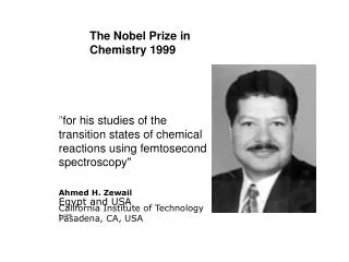
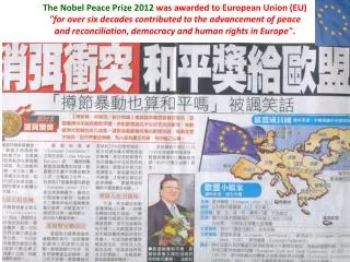
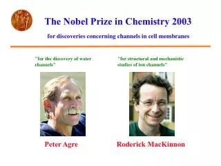
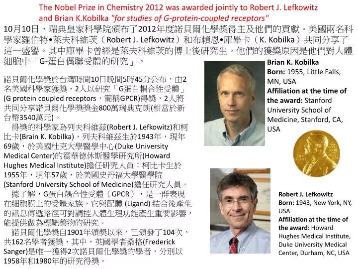
The Nobel Prize in Chemistry 2012 was awarded jointly to Robert J. Lefkowitz and Brian K.Kobilka "for studies of G-protein-coupled receptors".

E N D
The Nobel Prize in Chemistry 2012 was awarded jointly to Robert J. Lefkowitz and Brian K.Kobilka"for studies of G-protein-coupled receptors" 10月10日,瑞典皇家科學院頒布了2012年度諾貝爾化學獎得主及他們的貢獻。美國兩名科學家羅伯特•萊夫科維茨(Robert J. Lefkowitz)和布賴恩•庫畢卡(K. Kobilka)共同分享了這一盛譽。其中庫畢卡曾經是萊夫科維茨的博士後研究生。他們的獲獎原因是他們對人體細胞中「G-蛋白偶聯受體的研究」。 Brian K. Kobilka Born: 1955, Little Falls, MN, USA Affiliation at the time of the award: Stanford University School of Medicine, Stanford, CA, USA 諾貝爾化學獎於台灣時間10日晚間5時45分公布,由2名美國科學家獲獎,2人以研究「G蛋白耦合性受體」(G protein coupled receptors,簡稱GPCR)得獎,2人將共同分享諾貝爾化學獎獎金800萬瑞典克朗(相當於新台幣3540萬元)。 得獎的科學家為列夫科維茲(Robert J. Lefkowitz)和柯比卡(Brain K. Kobilka),列夫科維茲生於1943年,現年69歲,於美國杜克大學醫學中心(Duke University Medical Center)的霍華德休斯醫學研究所(Howard Hughes Medical Institute)擔任研究人員;柯比卡生於1955年,現年57歲,於美國史丹福大學醫學院(Stanford University School of Medicine)擔任研究人員。 據了解,G蛋白耦合性受體(GPCR),是一群表現在細胞膜上的受體家族,它與配體 (Ligand) 結合後產生的訊息傳遞路徑可對調控人體生理功能產生重要影響,能提供做為標靶藥物的研究。 諾貝爾化學獎自1901年頒獎以來,已頒發了104次,共162名學者獲獎,其中,英國學者桑格(Frederick Sanger)是唯一獲得2次諾貝爾化學獎的學者,分別以1958年和1980年的研究得獎。 Robert J. Lefkowitz Born: 1943, New York, NY, USA Affiliation at the time of the award: Howard Hughes Medical Institute, Duke University Medical Center, Durham, NC, USA
Academician David Ma's "Argonne’s connections to this year’s Chemistry Nobel Prize“ http://www.anl.gov/articles/advanced-photon-source-lights-way-2012-chemistry-nobel?utm_source=Argonne+Today&utm_campaign=1ef005c1f7-AT+12%2F10%2F11&utm_medium=email This is an image of a G-protein-coupled receptor signaling complex whose structure was identified in 2011. The receptor is in magenta while the different G protein subunits are colored green, red and blue. Stanford biochemist Brian Kobilka shared the 2012 Nobel Prize in Chemistry for his work in determining the structure of this activated GPCR using X-rays provided by Argonne’s Advanced Photon Source.
Figure 1. Cartoon of a cell with its interior (light blue) and exterior (blue), with their different chemical compositions separated by a phospholipidbilayer. The bilayer contains many proteins. Shown are two copies of a GPCR with specificity for a diffusible ligand (yellow). The fraction of receptors with bound ligand is governed by the ligand concentration. The receptor to the left is unoccupied and non-activated, and the receptor to the right is occupied by a ligand, bound to a G-protein (red), and activated. The ligand does not pass through the membrane; the signal is transmitted by conformational changes in the receptor protein.
Figure 2. Left: Ternary complex model adapted from (17). A thermodynamic cycle describes the formation of a complex of ligand (yellow), receptor (blue) and G-protein (red). Right: Crystal structure of an active ternary complex. Ribbon model drawn from the coordinates from file 3sn6.pdb (19) using the software Molmol (20).
Figure 3. The prediction of seven helices in βAR is shown above rhodopsin. The homologous amino acid sequences in helices five, six and seven are aligned, and identities and similarities are coloured. Adapted from reference 33.
Figure 4. Structural basis of the GPCR signalling mechanism. Non-activated βAR (2bar.pdb) is shown to the left, and activated βAR bound to ligand and G-protein (3sn6.pdb) to the right. At the top, the receptor in the membrane is drawn with blue ribbon that traces the backbone. The bottom view is from the inside of the cell membrane, with the receptor shown using a space-filling model with hydrophobic side chains in dark blue.