Download
1 / 32
320 likes | 331 Vues
Symptoms and Signs in Respiratory System Dr. Nawal N Binhasher Assistant professor, Medical Consultant department of medicine. History: . Symptoms: Cough Sputum Hemoptysis Dyspnea Chest pain (chest tightness) Wheezing. cough. Definition : cough reflex arc. Types :.

E N D
Symptoms and Signs in Respiratory SystemDr. Nawal N BinhasherAssistant professor, Medical Consultant department of medicine
History: Symptoms: Cough Sputum Hemoptysis Dyspnea Chest pain (chest tightness) Wheezing
cough Definition: cough reflex arc
Types: • Acute (< 3 wks) ex: RTI • Subacute (3-8 wks) ex: post RTI • Chronic (>8 wks) ex:bronchiectasis
Causes of acute cough: • Acute upper respiratory tract infection. • Acute lower respiratory tract infection (pneumonia). • Acute exacerbation of underlying chronic pulmonary disease. • Pulmonary Embolism (PE).
Causes of subacute cough: • Post-infection of upper or lower respiratory tract. • Angiotensin Converting Enzyme Inhibitors (ACE-I) medication.
Common causes of chronic cough usually with a normal CXR: • Upper airway cough syndrome (it is related to allergic, non-allergic or vasomotor rhinitis, naso-pharyngitis, & sinusitis. i.e postnasal drip «PND») • Bronchial Asthma • Gastroesophageal reflux disease
Other Respiratory Causes: • Chronic bronchitis (COPD, eosinophilic) • Bronchiectasis • Neoplasm • Interstitial lung disease (ILD) • Lung abscess • Obstructive sleep apnea (OSA) • Tracheobronchial foreign body or mass • Nasal polyps & others……
Non-Respiratory Causes: Mediastinal: • external tracheal compression ex: enlarged LN • Tumors, cysts, masses Cardiac: • LVF • Severe MS ENT: • Acute/chronic sinusitis • PND (perennial, allergic, or vasomotor rhinitis)
Cont’n: GI: • GERD • Esophageal dysmotility, stricture, or pouch • Esophago-bronchial fistula CNS: • CVA • MS • MND • Parkinson’s disease
Cont’n Drugs: • ACE-Inhibitors • Some inhaler preparations can cause cough Others: • Idiopathic • Ear wax (vagal nerve stimulation) • Psychogenic
Sputum: • Amount: N amount < 100mls of mucus/day • Color: N, clear & white mucus • Smell: N, not smelly Ex: chronic large amount of purulent sputum may suggest bronchiectasis while acute one may indicate lobar pneumonia. Ex: foul-smelling purulent sputum may indicate lung abscess with anaerobic infection Ex: pink frothy secretions occurs in pulmonary edema
Hemoptysis: • It’s a blood-stained sputum • Varies from streaks of blood to massive bleeding (>100 - 600mls /24 hrs) • It should be investigated thoroughly • Commonest cause is acute infection like exacerbation of copd but other serious causes should be rolled out • Other causes: PE, Bronchogenic ca., pul TB, bronchiectasis, lung abscess,
Cont’n • Pulmonary hemorrhage from any cause like: Goodpasture’s syndrome or rupture of a mucosal blood vessel after a vigorous coughing • Non-respiratory causes: CVS: severe MS, & acute LVF. Bleeding Diathesis should be excluded. • Rusty sputum (when purulent sputum is mixed with blood) eg: lobar pneumonia
Dyspnoea: • Defined as: experience of discomfort in breathing or an awareness of respiratory distress & physiologically its an ↑ in the level & work of breathing. • Onset: • Instantaneous: pneumothorax, PE • Min.s – hrs: * Aw disease: (BA, copd exacerbʼn, UAW obstrcʼn) * parenchymal disease: (pneumonia, pul hage, pul edema..) * pul vascular disease: (PE) * cardiac disease: ( MI,……. ) * metabolic acidosis * hyperventilation syndrome.
Cont’n: 3. Subacute (days): * Many of the above plus: * Pl. effusion * lobar collapse * Acute Interstitial pneumonia * SVC obstruct’n * Pul vasculitis 4. Chronic (months-years): * COPD & BA * Diffuse parenchymal dis: (IPF, sarcoidosis, bronchiectasis) * Hypoventilat’n:(neuromuscular weakness, chest wall defor) * Anemia * Thyrotoxicosis
Cont’n: • Severity (grading): Dyspnea can be graded from І – IV based on the NYHA classification. Chest pain: Pul causes of CP: 1. pul vasculature: • Acute PE • Pul HTN & Corpulmonale 2. Lung parenchyma: • Pneumonia • Cancer • sarcoidosis
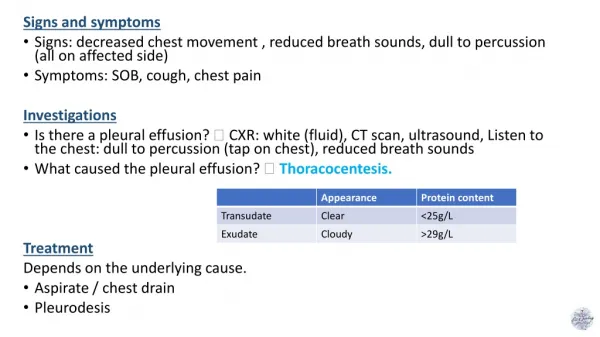
Cont’n: 3. Pleura & plural spaces: • Pneumothorax • Pleuritis & serositis • Pleural effusion 4. psychogenic/psychosomatic Wheezing: It’s a continuous whistling, not diagnostic for asthma & can occur in other resp diseases like copd.
Other symptoms: • Runny, blocked nose & sneezing: may occur in both common cold & allergic rhinitis (loss of smell = inosmia, runny nose = rhinorrhea) • Nocturnal fever may accompany TB, pneumonia, & mesothelioma. • Nocturnal sweating can occur in TB, lymphoma, & lung abscess. • Hoarseness may be secondary to laryngitis, VC tumor, & RLN palsy in apical lung CA. • Symptoms of corpulmonale (abd & ankle swelling, ….)
Other aspects of history: • Details of the respiratory system symptom should be inquired such as; onset, duration, character, radiation/severity/grading, frequency, aggravating & relieving factors, & associated symptoms. • PMH of a respiratory disease • Smoking history in details • Drug history including IV drug abuser (lung abscess) & alcohol consumption (aspiration pneumonia) • Inquiry about occupat’n & or previous jobs • Pets history
Clinical examination (signs): * General appearance * General system * Chest examination In general appearance, look for: • Respiratory distress {count RR, normal 14-20bpm Tachypnea = ↑ rate of breathing Hyperapnea = ↑ level of ventilation, and look to the accessory muscles; sternomastoids, scalene, platysma & strap muscles of neck & abdominal muscles, if they are in use?}
Cont’n • Coughing;character (bovine cough…) • Sputum; • Abnormal sound; stridor (croaking noise, loudest on inspiration 2°to larynx, trachea or large airways obstruction), or wheezing. • Abnormal voice; hoarseness • Surroundings; likecontainers of sputum, O2 mask, IV lines or medications respiratory aids or machines..
General system examination: • Hands: • Clubbing (check respiratory causes) • Tar staining • Weakness of hand’s small muscles (abduction) • Wrist: • Pulse: rate & character • Flapping tremors (asterixis) • BP: pulsus paradoxux (asthma), hypotension
Cont’n • Neck: • JVP: ↑ in corpulmonale & SVC obstruct’n but not pulsatile. • LN: enlargement in CA bronchus or mets • Face: • Eye: Horner’s syndrome in CA bronchus • Tongue: central cyanosis • SVC obstruction: plethoric & cyanosed, periorbital edema, injected conjuctvae & +ve Pemberton’s sign
Chest examination: • Inspection: • Shape: AP diameter compared to transverse (barrel-chest), pectus excavatum, pectus carinatum, kyphoscoliosis,…. others • Symmetry: assessment of upper & lower lobes should be done posteriorly looking for ↓ or delayed chest movement during moderate respirat’n. • Scars: from previous operat’n or chest drains or cautery marks or radiotherapy markings. • Prominent veins: in case of SVC obstruct’n
Palpation: • Trachea: normally central, slight Rt displacement could be N. Check for gross displacement. Tracheal tug means the N distance bet sternal notch & cricoid cartilage is < 3-4 finger breadths & occurs in chest overexpansion as copd. • Apex beat & mediastinum: Check for displacement. • Chest expansion: N expansion ≥ 5cm • Tactile vocal fremitus (TVF): can be done with the palm of one hand.
Percussion: • Should be done symmetrically (Lt compared with the Rt), posteriorly (the back), anteriorly (the front) & laterally (the sides). • Supraclavicular area, then clavicles should be percussed directly to evaluate the upper lobes. • Liver dullness: of the upper edge starting at the 6th rib MCL, resonant note below this area indicates hyper-inflation (copd, severe asthma) • Cardiac dullness: may be ↓ in hyperinfated chest.
Auscultation: Using the diaphragm of a stethoscope & comment on the following: • Breath sounds (BS): • Intensity: N or ↓ as in (consolidation, collapse, pl effusion, pneumothorax, lung fibrosis) • Quality: Vesicular or bronchial in consolidation • Differentiation between vesicular & bronchial BS: Vesicular: louder &longer on inspiration than expiratory phase & has no gap between the 2 phases Bronchial: louder &longer on exp phase & has a gap between the 2 phases
2. Added Sounds: • Type: Wheezes or Crackles or friction rub • Timing: inspiratory or expiratory • Wheezes: are continuous musical polyphonic sound, heard louder on expiration & can be heard on inspiration which may imply severe AW narrowing. High pitched- wheezes are found in BA due to acute/chronic airflow limitation & low pitched in copd. Localized monophonic wheeze due to fixed AW obstruct’n in CA bronchus. • Crackles: interrupted non-musical inspiratory sound • Crackles may be early, late or pan-inspiratory & fine, medium or coarse. Ex: late/pan-insp coarse crackles in bronchiectasis, late/pan-insp medium crackles in pul edema , late/pan-insp fine crackles in pul fibrosis
friction rub: It’s due to thickened or roughened pl surfaces rub together as lungs expand & contract & give off a continuous or intermittent grating sound. It indicates pleurisy & may be heard in pneumonia or pul infarction. • Vocal Resonance: • It’s the ability to transmit sounds. • Ask patients to say 44 (Arabic) or 99 (English) & listen for the transmitted sound which may be ↓ or ↑ or N (low pitched component of speech heard with booming & high pitched become attenuated).
4. Egophony: When the patient with consolidation is asked to say ‘e’ it sounds like ‘a’ • Whispering pectoriloquy: The whispered speech is heard very loudly over the consolidated area. Other signs should be looked for to complete the respiratory system examination “signs of complications” • Signs of pul HTN or corpulmonale. 2. Signs of SVC obstruction. 3. Signs of CA bronchus mets, or extension
Secondary pul HTN or corpulmonale: • Should be suspected in: • Chronic airflow limitation such as copd • Pulmonary fibrosis • Chronic pul thromboembolism • OSA • Severe kyphoscoliosis/marked obesity • Signs: loud P2 of S2 + signs of RHF Thank you Any ?