Download
1 / 1
10 likes | 75 Vues
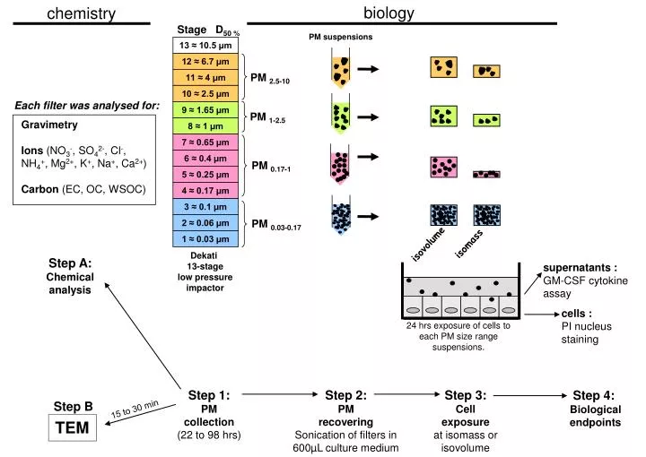
biology. chemistry. 13 ≈ 10.5 µm. 12 ≈ 6.7 µm. 11 ≈ 4 µm. ●. ●. ●. ●. ●. ●. ●. 10 ≈ 2.5 µm. ●. ●. ●. ●. ●. ●. ●. ●. 9 ≈ 1.65 µm. 8 ≈ 1 µm. 7 ≈ 0.65 µm. 6 ≈ 0.4 µm. 5 ≈ 0.25 µm. 4 ≈ 0.17 µm. 3 ≈ 0.1 µm. 2 ≈ 0.06 µm. 1 ≈ 0.03 µm. Stage D 50 %. PM suspensions.

E N D
biology chemistry 13 ≈ 10.5 µm 12 ≈ 6.7 µm 11 ≈ 4 µm ● ● ● ● ● ● ● 10 ≈ 2.5 µm ● ● ● ● ● ● ● ● 9 ≈ 1.65 µm 8 ≈ 1 µm 7 ≈ 0.65 µm 6 ≈ 0.4 µm 5 ≈ 0.25 µm 4 ≈ 0.17 µm 3 ≈ 0.1 µm 2 ≈ 0.06 µm 1 ≈ 0.03 µm Stage D50 % PM suspensions PM 2.5-10 Each filter was analysed for: PM 1-2.5 Gravimetry Ions (NO3-, SO42-, Cl-, NH4+, Mg2+, K+, Na+, Ca2+) Carbon (EC, OC, WSOC) ● ● ● ● ● PM 0.17-1 ● ● ● ● ● ● ● ● ● ● ● PM 0.03-0.17 isomass isovolume Dekati 13-stage low pressure impactor Step A: Chemical analysis supernatants : GM-CSF cytokine assay cells : PI nucleus staining 24 hrs exposure of cells to each PM size range suspensions. Step 1: PM collection (22 to 98 hrs) Step 2: PM recovering Sonication of filters in 600µL culture medium Step 3: Cell exposure at isomass or isovolume Step 4: Biological endpoints Step B 15 to 30 min TEM