Download
1 / 40
1.35k likes | 2.98k Vues
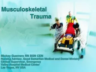
Musculoskeletal Trauma. Silvia De La Guardia RN, BSN, CEN Tenet Health Systems Emergency Department. Learning Objectives. List three signs or symptoms of joint dislocations. Identify how much blood can accumulate in the femur compartment.

E N D
MusculoskeletalTrauma Silvia De La Guardia RN, BSN, CEN Tenet Health Systems Emergency Department
Learning Objectives • List three signs or symptoms of joint dislocations. • Identify how much blood can accumulate in the femur compartment. • Explain why prompt reduction of joint injuries is important. • Identify signs of a femur and pelvic fracture. • Identify injuries or complications associated with pelvic fractures. • Discuss emergent management of compartment syndrome.
Musculoskeletal Trauma • Musculoskeletal injuries often appear dramatic, rarely cause immediate threat to life or limb • Major injuries, especially to long bones, indicate significant forces applied to body • Imperative to recognize and control hemorrhage from musculoskeletal injures in primary survey • Suspect neurovascular injury with any injury to the bones of an extremity. • Splinting significantly decreases bleeding • Reducing motion • Tamponade effect
Mechanism of Injury • Motor Vehicle Crash • Patella injury • Fractured femur • Hip fracture/dislocation • Assault • Fall • Calcaneus fracture • Compressed vertebral bodies • Sports • Recreational and home activities
Assessment Inspection • Appearance of Extremity • Normal vs. abnormal • Difference between injured and non-injured • Integrity of injured area • Deformity - Burns • Contusions - Tenderness • Abrasions - Lacerations • Punctures - Swelling • Bleeding - Protrusion of bone
Assessment Palpation • Six P’s • Pulse • Pain • Pallor • Paresthesias • Paralysis • Pressure • Palpate for stability and crepitus Circulation Pulse Cap refill Temp/color Swelling Motor Paralysis Sensation Pain Paresthesia
Pathophysiologic Changes BLOOD LOSS • Large blood loss is due to damage to arteries or veins close to bones • Edema can cause compression of structures. • Physiological changes activated to minimize damage: • Clotting system activation to decrease bleeding • Cell membrane restoration to enhance fluid reabsorption • Increased collateral blood flow to promote healing
Pathophysiologic Changes • Bone/joint displacement can compress surrounding vessels and nerves • As arterial blood is obstructed, tissue oxygenation ceases. Ischemia/tissue death. • In this process: • Pain increases - Pale limb • Weak pulses - Skin cyanotic • Cool skin - Increased capillary refill
Pathophysiologic Changes NEUROLOGIC CHANGES • Nerves can be compressed or lacerated • Nerve injury results in • Diminished pain • Partial or complete loss of motor • Partial or complete loss of sensation
Pathophysiologic Changes FRACTURES • Open - Closed • Complete - Incomplete • Comminuted - Greenstick • Impacted - Displaced • Fissure - Spiral
TYPES OF INJURIES Blunt injures • Acceleration/deceleration mechanism • Falls, MVC, work/home injuries • Assaults, sport injuries • Include • Fractures, dislocations, • Strains: Injury to muscle or tendon • Sprains: Injury to ligament • Ligamentous tears Penetrating Injuries • Penetrating objects • Open fractures
Joint Injuries • Considered limb-threatening injuries • Joint dislocations • Normal range of motion is exceeded • Complicated by neurovascular compromise • Associated fractures • Avascular necrosis • Complication of joint injuries • Caused by delay reduction • Knee dislocations require immediate reduction to prevent complications • Peroneal nerve injury • Popliteal artery disruption
Joint Injuries Signs and Symptoms • Inability to move the affected area • Neurovascular compromise • Abnormal range of motion • Pain • Deformity of joint • Edema
Femur Fractures • Result from major trauma • Common mechanism of injury • Falls • MVC • penetrating wounds (GSW) • Femoral neck fractures common • Femur fractures may not be obvious • Result in significant blood loss
Femur Fractures Signs and Symptoms • Pain • Inability to bear weight • Shortened extremity • Internal or external rotation • Edema or deformity • Hypovolemic shock
Pelvic Fractures Classification • Stable: • Pelvic ring fracture in one place • Stable to palpation • Can withstand normal physiologic forces • Without abnormal deformation • Unstable • Pelvic ring fractured in more than one place • Bony instability with palpation
Pelvic Fractures • Can be classified by type of force • External rotation (anteroposterior) • Lateral compression • External rotation (abduction) • Shear • These can be life-threatening and often have large blood loss and genitourinary injuries. • Bleeding significant enough to cause hypovolemic shock
Pelvic Fractures • Injuries to pelvis can be: • Open: associated with injuries to perineum, rectum, genitourinary system. Have higher mortality rate. • Closed.
Pelvic Fractures Signs and Symptoms • Intra-abdominal or genitourinary injury • Look for blood in meatus • Higher mortality rate • Shortening and abnormal rotation of the affected leg • Pain to pelvic area, back, thighs • Hypovolemic shock
Interventions • Stabilize pelvic fractures • PASG • Wrap pelvis in sheet • External fixator • Assist with additional diagnostic studies • Radiographs • CT scan • Cystogram • Angiogram • Embolization
Open Fractures • Considered Limb-threatening injuries • Consider contaminated avoid retracting bone back into tissue • Higher risk of infection • Poor wound healing • Osteomyelitis • Sepsis
Open Fractures Signs and Symptoms • Skin disruption • Protrusion of bone • Pain • Bleeding can be minimal or severe • Neurovascular compromise • Do not move fracture: minimize more damage • Call orthopedic consult early • Realign to restore blood flow and nerve integrity • Operative debridement
Emergent Interventions • Obtain wound culture • Irrigate the wound with sterile saline • Cover open wounds with dry sterile dressing • Administer medications • Tetanus • Antibiotics • Inspect dressings frequently
Avascular Injury • Acutely avascular extremity must be recognized promptly and treated emergently • Assess temperature, color, capillary refill, pulses • Early operative revascularization is critical • Muscles and nerves do not tolerate lack or arterial blood flow for longer than 6 hours • Use of tourniquets is controversial • Indicated for ongoing hemorrhage uncontrolled by direct pressure • Use of tourniquets is a “Life over limb” decision • Risk of tourniquet increase with time
Amputation • Traumatic event for patient: • Physically and emotionally • Amputations may benefit from a tourniquet • Replantation • Injury of an isolated extremity • Clean sharp amputations • Fingers • Distal extremity, below the knee or elbow • Wrist, forearm • Pediatric patient
Amputation Signs and Symptoms • Obvious tissue loss • Pain • Evidence of hypovolemic shock • Bleeding (minimal to severe) • Complete: less bleedingthan partial because of retraction of arteries. • Avulsive type complete amputation can result in severe bleeding.
Emergent Interventions • Control active bleeding • Remove gross debris • Elevate and splint the stump • Place amputated part on ice bath: do not freeze • Wrap in saline soaked gauze • Place in plastic bag then • Place bag on ice or ice water mixture • Prepare patient for admission or transfer • Administer medications: pain meds, antibiotics.
Crush Injuries • Life-threatening and difficult to treat • Cellular destruction • Damage to vessels • Result from • prolonged entrapment • Crushing blow • Sequelae • Hemorrhage and fluid loss • Destruction of muscle • Infection • Compartment syndrome • Rhabdomyolysis
Crush Injuries Sign and Symptoms • Massively crushed pelvis or extremities • Soft tissue swelling • Pain • Hypovolemic shock • Compartment syndrome • Loss of neurovascular function distal to injury
Emergent Interventions • Administer intravenous fluids to increase urinary output and excrete myoglobin • Elevate the injured extremity • Cleanse open wounds • Reassess urinary output • Motor and sensory function • Prepare for surgical debridement
Compartment Syndrome • Pressure increases inside fascial compartment Decreased perfusion and cellular ischemia. • Nerves, vessels, muscles can be compressed. • Prolonged ischemia results in • Pain • Necrosis • Loss of function • Amputation • Degree of damage • Amount of pressure • Time
Compartment Syndrome • Internal sources: • Bleeding • Tissue edema • Crush injuries • External sources • Tight dressings • Cast, excessive traction • More frequently seen • Lower leg • Forearm
Compartment Syndrome Signs and Symptoms • Pulses • Loss of pulse uncommon • Late unreliable sign • Pain • Disproportionate to injury • With passive movement • Pallor: Poor color, cool temperature • Paresthesia: Numbness, tingling, • Paralysis: motor dysfunction, injury to nerves, weak. • Pressure: compartment will feel tense, swollen
Emergent Interventions • Remove dressings, cast and splints • Elevate limb to level of the heart • Assist with measurement of compartment pressures • Reassess neurovascular status • Surgical consultation: Fasciotomy (may prevent muscle/neurovascular damage and loss of limb)
Diagnostics • X-Rays • Different views (anterior, posterior, lateral) • Film should include joints above/below. • Angiography • Indicated to identify tears or compressions in arteries/veins of extremity.
Interventions • Control bleeding • Splint and immobilize affected extremity • Apply ice • Elevate extremity: compartment syndrome keep at level of heart • Administer medication: antibiotics, tetanus, pain • Prepare for definitive stabilization • Provide psychosocial support • Prepare for operation, admission, or transfer, discharge
Evaluation • Monitor breathing effectiveness and rate of respiration. • Tachypnea, rales, and wheezes may be indicators of fat embolus syndrome. • Reassess and document 5 P’s.
Learning Objectives • List three signs or symptoms of joint dislocations. • Identify how much blood can accumulate in the femur compartment. • Explain why prompt reduction of joint injuries is important. • Identify signs of a femur and pelvic fracture. • Identify injuries or complications associated with pelvic fractures. • Discuss emergent management of compartment syndrome.
References • ATLS, advanced trauma life support for doctors (8th ed.). (2008). Chicago, IL: American College of Surgeons. • Beers, M. H. (2006). The Merck manual of diagnosis and therapy (18th ed.). Whitehouse Station, N.J.: Merck Research Laboratories. • Coven, D. L., & Yang, E. H. (n.d.). Medscape.com. Retrieved September 1, 2010, from emedicine.medscape.com/article/1910735-overview • Howard, P. K., Steinmann, R. A., & Sheehy, S. B. (2010). Sheehy's emergency nursing: principles and practice. (6th ed.). St. Louis, Mo.: Mosby Elsevier. • Tintinalli, J. E., & Stapczynski, J. S. (2011). Tintinalli's emergency medicine: a comprehensive study guide (7th ed.). New York: McGraw-Hill. • TNCC: trauma nursing core course (5th ed.). (2000). Park Ridge, Ill.: Emergency Nurses Association.