Download
1 / 123
1.31k likes | 1.67k Vues
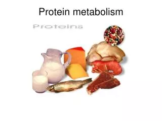
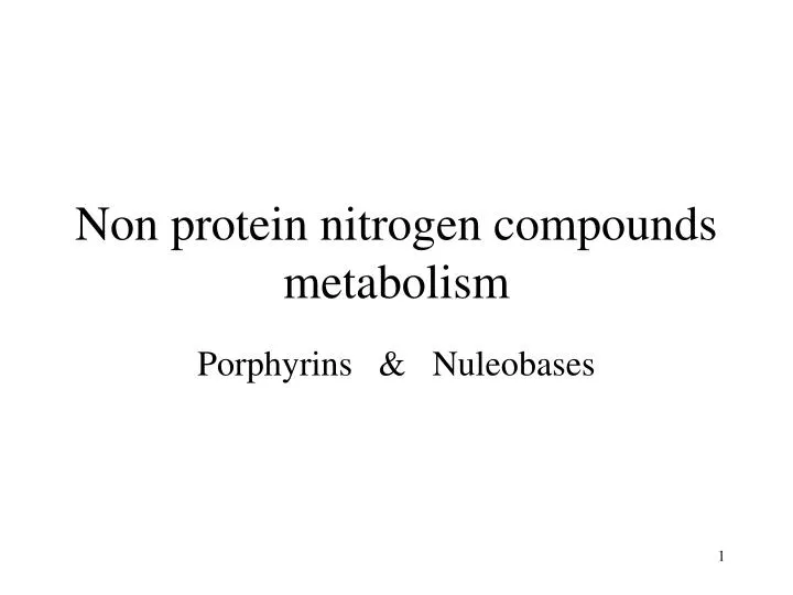
Non protein nitrogen compounds metabolism. Porphyrins & Nuleobases. Heme Metabolism. Heme biosynthesis Heme degradation. Biosynthesis of Heme. Production of Aminolevulinic acid from 2 carbon amino acid glycine and succinyl CoA in the presence of Ala synthase

E N D
Non protein nitrogen compounds metabolism Porphyrins & Nuleobases
Heme Metabolism • Heme biosynthesis • Heme degradation
Biosynthesis of Heme • Production of Aminolevulinic acid from 2 carbon amino acid glycine and succinyl CoA in the presence of Ala synthase • Requires two vitamines - pyridoxal phosphate and pantothenic acid • ALA synthase is an important rate limiting factor (heme represses - sex hormones enhance - high glucose blocks)
Two ALA molecules are joined in the presence of the enzyme delta aminolevulinic dehydratase • Forms porphobilinogen • Lead inhibits this step
Four porphobilinogen molecules condense to form hydroxymethylbilane and then uroporphyrinogen III • Requires porphobilinogen deaminase (uroporphyrinogen synthtase) and uroporphyrinogen III co-synthtase
Decarboxylation (remove COOH) of the four acetic acid side chains of uroporphyrinogen III to form methyl (CH3) • Forms coproporphyrinogen III • Catabolized by the enzyme uroporphyrinogen decarboxylase
Conversion of coproporphyrinogen III to protoporphyrinogen III • Two propionic acid (CH2-CH2-COOH) convert to two vinyl (CH2=CH2) • Requires coproporphyrinogen oxidase and oxygen as a hydrogen acceptor • Moves heme synthesis back into the mitochondria
Fifteen possible isomers of protoporphyrinogen can form • Normal mitochondrial physiology leads to the formation of only one of these isomers (protoporphyrinogen IX) • Protoporphyrinogen oxidase is involved in this reaction and oxygen as a hydrogen acceptor
Heme • A complex of iron and protoporphyrin (a porphyrin ring)
Porphyrins • Protoporphyrin • Coproporphyrin • Uroporphyrin
COORDINATED REGULATION OF HEME AND GLOBIN SYNTHESIS: • Heme: • inhibits activity of pre-existing -ALA synthase • diminishes the transport of -ALA synthase from cytoplasm to mitochondria after synthesis of the enzyme. • represses the production of -ALA synthase by regulating gene transcription. • stimulates globin synthesis to ensure that levels of free heme remain low in concentration. Inhibition of the synthase and stimulation of globin synthesis are the most important aspects in balancing hemoglobin production.
Heme Biosynthesis: Porphyrias • Cruelly referred to as a Vampire’s disease. • Can be caused by lead poisoning: The fall of the Roman Empire!
Not a ‘vampire’s’ disease Some symptoms of porphyrias have lead people to believe that these diseases provide some basis for vampire legends: • Extreme sensitivity to sunlight • Anemia This idea has been discarded both for scientific reasons: • Porphyrias do not cause a craving for blood. • Drinking blood would not help a victim of porphyria. And for compasionate reasons:Porphyria is a rare, but frightening condition: hard to diagnose and there is no cure.
PORPHYRIAS Mitochondria GLYCINE + SuccinylCoA Agent Orange ALA synthase 3p21/Xp11.21 d-aminolevulinic acid(ALA) ALA-dehydratase Deficiency porphyria ALA dehydratase 9q34 Porphobilinogen(PBG) Acute intermittent porphyria PBG deaminase 11q23 hydroxymethylbilane Congenital erythropoietic porphyria Uroporphyrinogen III cosynthase 10q26 uroporphyrinogen III Uroporphyrinogen decarboxylase Prophyria cutanea tarda 1q34 coprophyrinogene III Coproporphyrinogen oxidase Herediatary coproporphyria 9 Protoporphyrinogene IX Protoporphyrinogen oxidase Variegate porphyria protoporphyrin IX 1q14 Ferrochelatase Erythropoietic protoporphyria Heme 18q21.3
NADPH NADPH NADP+ NADP+ Heme Degradation Heme Catabolism HEME O2 (opens the porphyrin ring) Fe+3 BILIVERDIN BILIRUBIN BILIRUBIN diglucuronide BILE
Stercobilin excreted in feces Urobilin excreted in urine Hemoglobin Globin Urobilinogen formed by bacteria Heme O2 Heme oxygenase CO Biliverdin IX NADPH Bilirubin diglucuronide (water-soluble) Biliverdin reductase NADP+ 2 UDP-glucuronic acid Bilirubin (water-insoluble) Bilirubin (water-insoluble) via blood to the liver BLOOD CELLS KIDNEY reabsorbed into blood INTESTINE via bile duct to intestines LIVER Figure 2. Catabolism of hemoglobin
Jaundice Hyperbilirubinemia: Two forms: Direct bilirubin: Conjugated with glucoronic acid Indirect bilirubin: unconjugated, insoluble in water.
What’s the cause of jaundice? • 1- Increased production of bilirubin by hemolysis or blood disease: • Increase in blood indirect bilirubin • Called pre-hepatic jaundice • Stool color remains normal. • 2- Abnormal uptake or conjugation of bilirubin: • Leads to non-hemolytic unconjugated hyperbilirubinemia • Increased indirect bilirubin. • Stool color turns gray. • Caused by liver damage or disease.
3- Cholestasis = Problems with bile flow. • a: Intrahepatic cholestasis: hyper conjugated bilirubinemia • Increase in blood indirect and direct bilirubin • Caused by liver damage or disease: eg cirrhosis, hepatitis • Can also occur in pregnancy: • b:Extrahepatic cholestasis: • Blockage of bilirubin transport in the bilary tract. • Increased direct bilirubin. • Stool color turns gray. • Caused by: Tumors or gall stones.
Examples of hyperbilirubinemia A. Hemolytic anemia C. Biliary duct stone B. Hepatitis excess hemolysis unconjugated bilirubin (in blood) conjugated bilirubin (released to bile duct) unconjugated bilirubin (in blood) conjugated bilirubin (in blood) unconjugated bilirubin (in blood) conjugated bilirubin (in blood)
Roles of Nucleotides • Precursors to nucleic acids (genetic material and non-protein • enzymes). • Currency in energy metabolism (eg. ATP, GTP). • Carriers of activated metabolites for biosynthesis • (eg. CDP, UDP). • Structural moieties of coenzymes (eg. NAD, CoA). • Metabolic regulators and signal molecules (eg. cAMP, • cGMP, ppGpp).
Nitrogenous Bases Purines Pyrimidines N1: Aspartate Amine C2, C8: Formate N3, N9: Glutamine C4, C5, N7: Glycine C6: Bicarbonate Ion
Purine degredation AMP deamination in muscle, hydrolysis in other tissues. Xanthine oxidase:contains FAD, molybdenum, and non-heme iron. In primates, uric acid is the end product, which is excreted.
Purine Nucleotides • Get broken down into Uric Acid (a purine)
Common treatment for gout: allopurinol Allopurinol is an analogue of hypoxanthine that strongly inhibits xanthine oxidase. Xanthine and hypoxanthine, which are soluble, are accumulated and excreted.
Purine degredation in other animals
Uric Acid Excretion • Humans – excreted into urine as insoluble crystals • Birds, terrestrial reptiles, some insects – excrete isoluble crystals in paste form (conserve water) • Others – further modification : Uric Acid Allantoin Allantoic Acid Urea Ammonia
Impaired excretion or overproduction of uric acid Uric acid crystals precipitate into joints (Gouty Arthritis), kidneys, ureters (stones) Lead impairs uric acid excretion – lead poisoning from pewter drinking goblets Fall of Roman Empire? Xanthine oxidase inhibitors inhibit production of uric acid, and treat gout Allopurinol treatment – hypoxanthine analog that binds to Xanthine Oxidase to decrease uric acid production Gout
Catabolism of pyrimidines
Biosynthetic routes: De novo and salvage pathways De novo pathways Almost all cell types have the ability to synthesize purine and pyrimidine nucleotides from low molecular weight precursors in amounts sufficient for their own needs. The de novo pathways are almost identical in all organisms. Salvage pathways Most organisms have the ability to synthesize nucleotides from nucleosides or bases that become available through the diet or from degredation of nucleic acids. In animals, the extracellular hydrolysis of ingested nucleic acids represents the major route by which bases become available.
Reutilization and catabolism of purine and pyrimidine bases blue-catabolism red-salvage pathways endonucleases: pancreatic RNAse pancreatic DNAse phosphodiesterases: usually non-specific
Purine Catabolism and Salvage • All purine degradation in animals leads to uric acid • Ingested nucleic acids are degraded by pancreatic nucleases, and intestinal phosphodiesterases in the intestine • Group-specific nucleotidases and non-specific phosphatases degrade nucleotides into nucleosides • Direct absorption of nucleosides • Further degradation Nucleoside + H2O base + ribose (nucleosidase) Nucleoside + Pi base + r-1-phosphate (n. phosphorylase) NOTE: MOST INGESTED NUCLEIC ACIDS ARE DEGRADED AND EXCRETED.
Intracellular Purine Catabolism • Nucleotides broken into nucleosides by action of 5’-nucleotidase (hydrolysis reactions) • Purine nucleoside phosphorylase (PNP) • Inosine Hypoxanthine • Xanthosine Xanthine • Guanosine Guanine • Ribose-1-phosphate splits off • Can be isomerized to ribose-5-phosphate • Adenosine is deaminated to Inosine (ADA)
Intracellular Purine Catabolism • Xanthine is the point of convergence for the metabolism of the purine bases • Xanthine Uric acid • Xanthine oxidase catalyzes two reactions • Purine ribonucleotide degradation pathway is same for purine deoxyribonucleotides
PRPP: a central metabolite in de novo and salvage pathways PRPP synthetase Enzyme inhinited by AMP, ADP, and GDP. In E. coli, expression is repressed by PurR repressor bound to either guanine or hypoxanthine. Roles of PRPP: his and trp biosynthesis, nucleobase salvage pathways, de novo synthesis of nucleotides
Example of a salvage pathway: guanine phosphoribosyl transferase In vivo, the reaction is driven to the right by the action of pyrophosphatase Shown: HGPRT, cells also have a APRT.
Purine Salvage • Adenine phosphoribosyl transferase (APRT) Adenine + PRPP AMP + PPi • Hypoxanthine-Guanine phosphoribosyl transferase (HGPRT) Hypoxanthine + PRPP IMP + PPi Guanine + PRPP GMP + PPi (NOTE: THESE ARE ALL REVERSIBLE REACTIONS) AMP,IMP,GMP do not need to be resynthesized de novo !
De novo biosynthesis of purines: low molecular weight precursors of the purine ring atoms
Synthesis of IMP The base in IMP is called hypoxanthine Note: purine ring built up at nucleotide level. precursors: glutamine (twice) glycine N10-formyl-THF (twice) HCO3 aspartate In vertebrates, 2,3,5 catalyzed by trifunctional enzyme, 6,7 catalyzed by bifunctional enzyme.