Download
1 / 55
570 likes | 835 Vues
CASE MANAGEMENT, PRESENTATION, DISCUSSION AND SHARING OF INFORMATION ON COMPLICATED SKIN AND SOFT TISSUE INFECTION. JONATHAN R. MALABANAN,MD DEPARTMENT OF SURGERY OSPITAL NG MAYNILA. C.S. 63M PANDACAN, MANILA. CHIEF COMPLAINT. Swelling, scrotal area. HISTORY OF PRESENT ILLNESS.

E N D
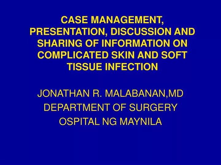
CASE MANAGEMENT, PRESENTATION, DISCUSSION AND SHARING OF INFORMATION ON COMPLICATED SKIN AND SOFT TISSUE INFECTION JONATHAN R. MALABANAN,MD DEPARTMENT OF SURGERY OSPITAL NG MAYNILA
C.S. 63M PANDACAN, MANILA
CHIEF COMPLAINT Swelling, scrotal area
HISTORY OF PRESENT ILLNESS 10 DAYS PTC SWELLING AND ERYTHEMA OF THE SCROTAL AREA PAIN AND TENDERNESS CRUSTING SKIN LESIONS (-) CONSULT OMMC OPD 1 DAY PTC INCREASED SEVERITY OF SWELLING , ERYTHEMA, PAIN AND TENDERNESS FEVER AND CHILLS (+) CONSULT OMMC SURGERY ER
PAST MEDICAL HISTORY - HPN X 20YRS, Metoprolol 50 mg tab BID - (+) DM • FAMILY HISTORY - UNREMARKABLE • PERSONAL/SOCIAL HISTORY - SMOKER, 25 pack years -NON- ALCOHOLIC BEVERAGE DRINKER
PHYSICAL EXAMINATION GENERAL: CONSCIOUS, COHERENT, NICRD BP: 130/90 CR= 90 RR=24 T=38.9 WT= 57 kg HEENT: PINK PALPEBRAL CONJUNCTIVA, ANICTERIC SCLERA, NO TPC, (-) CLAD, CHEST/LUNGS: SCE, NO RETRACTIONS, CLEAR BREATH SOUNDS HEART: ADYNAMIC PRECORDIUM, NRRR, NO MURMUR
PHYSICAL EXAMINATION >Perineum: (+) erythematous swelling scrotal area crusting skin lesion of the scrotum Tenderness Foul smelling d/c DRE: (+) hemorrhoidectomy site good sphincteric tone, tenderness Right anterolateral area
SALIENT FEATURES • 63/M, DM 2. (+) erythematous swelling on the scrotal area, crusting skin lesion of the scrotum Tenderness Foul smelling d/c 3. FEVER AND CHILLS
SCROTAL MASS NON-INFLAMMATORY NON-INFLAMMATORY INFLAMMATORY INFLAMMATORY TUMOR TUMOR RUBOR DOLOR MALIGNANT MALIGNANT BENIGN BENIGN TUMOR CALLOR
INFLAMMATORY UNCOMPLICATED COMPLICATED SKIN -EPIDERMIS -DERMIS FOLLICULITIS FURUNCLE SSS SUBCUTANEOUS TISSUE SUBCUTANEOUS ABSCESS CELLULITIS NECROTIZING FASCITIS FASCIA PYOMYOSITIS MYONECROSIS MUSCLE MYOSITIS
DO I NEED A PARACLINICAL DIAGNOSTIC PROCEDURE? NO. • I AM QUITE CERTAIN OF MY DIAGNOSIS • IT WILL NOT CHANGE MY TREATMENT PLAN
GOALS OF TREATMENT • RESOLUTION OF INFECTION • PREVENT RECURRENCE OF INFECTION
PRE-TREATMENT PREPARATION • PSYCHOSOCIAL SUPPORT • SCREENING FOR MEDICAL PROBLEMS: - OPTIMIZE PHYSICAL CONDITION OF THE PATIENT - ADEQUATE HYDRATION - ANALGESICS FOR PAIN AND FEVER
TREATMENT PLAN: Emergency Radical Wound Debribement Parenteral Antibiotic with coverage involving Streptococcus pyogenes benzylpenicillin 100,000 IU/kg i.v. every 6 hours And Metronidazole 500 mg Q8 Tetanus Prophylaxis 6,000 units TIM ( ) ANST Tetanus toxoid .5 ml TIM
OPERATIVE TECHNIQUE • Patient supine under SAB • Asepsis, Antisepsis • Sterile drapes placed • Longitudinal scrotal incision carried down from skin to dartos fascia • Intraop Findings noted
Intraop Findings • Necrotic tissue • Skin • Subq • Fascia • Normal tissue overlying necrotic tissue over the perineal area • Foul smelling discharge
Radical debridement done • NSS Flushing • Correct Instrument, Needle, and Sponge Count • Wet to Dry sterile dressing
TREATMENT • Radical Wound Debribement • MEDICAL: • Penicillin G 5 M units TIV q6 • Metronidazole 500 mg Q8 • Ketorolac 75 mg TIV q8 for pain and swelling • Intermediate Insulin
POST-TREATMENT DIAGNOSIS Fournier’s Gangrene S/P Radical Wound Debribement
Course in the Ward • IV antibiotics given • Adequate pain control • 6 hours post op • Arrested
POST-TREATMENT CARE • SUPPLY THE BASIC NEEDS OF THE PATIENT • COMFORT • ANALGESICS • MEDICATIONS – ANTIBIOTICS • ADEQUATE SUGAR CONTROL • SUPPORT ORGAN FUNCTION • MONITORING FOR COMPLICATIONS • TISSUE COVERAGE • ADVICE ON • HOME CARE • FOLLOW-UP PLAN
Fournier’s gangrene • Polymicrobial necrotizing fasciitis of the perineal- 21% perirectal-33% genital area-45% that extend rapidly along fascial planes to involve the groin, thighs, and abdominal wall
Epidemiology • According to CDC, 3/10,000 patient’s are diagnosed with Fournier’s Gangrene • Sex: M:F of 10:1 • Age: Mostly <3 months and aged 30- 60 y.o.
Predisposition toDisease Diabetes Mellitus- 60% Chronic Alcoholism-25-50% Immunosuppression Cigarette Smoking
Etiology and Pathogenesis • Polymicrobial infection • Mixed aerobic and anaerobic bacteria • Escherichia coli, Bacteroides, Clostridia, Staphylococci, Enterococci, Proteus and Pseudomonas
Common colorectal sources of infection perirectal, perianal, ischiorectal abscess perforation due to inflammatory disease neoplastic disease, instrumentation or trauma
Regardless of portal of entry • Local infection causes marked inflammatory reaction that extends to the deep fascial planes • Progresses to obliterative endarteritis and local ischemia
Direction of disease spread determined by the attachments of various fascial planes in the pelvis and perineum • Testes, epidydymes, bladder, and rectum are spared from necrosis because of the separate nonperineal blood supply
Prognosis • Mortality rates ranging from 20-25% • High morbidity exists among survivors • M/M increase with advanced age primary anorectal infection delayed treatment
shock or sepsis at presentation renal failure greater disease extent DM immunosuppression
Management • True surgical emergency that is rapidly progressive and potentially lethal • Once recognized: urgent surgical intervention with aggressive resuscitation • High dose parenteral antibiotics
Broad spectrum antibiotics ampisulbactam, ticarcillin/clavulanate, piperacillin/tazobactam, or imipinem, penicilinase-resistant, synthetic penicillin plus clindamycin plus antipseudomonal aminoglycoside
SSSS • Staphylococcal scalded-skin syndrome (SSSS) occurs predominantly in neonates and is caused by an exotoxin from phage group II S aureus • Punch biopsy with frozen section is useful since the cleavage plane in SSSS is the stratum corneum
NECROTIZING FASCIITIS • Necrotizing fasciitis is a deep-seated infection of the subcutaneous tissue that results in the progressive destruction of fascia and fat • Predisposing factors for the development of necrotizing fasciitis due to Strep. pyogenes include varicella ,penetrating injuries, minor cuts, burns, splinters, surgical procedures, childbirth, blunt trauma, and muscle strain
NECROTIZING FASCIITIS • Type I necrotizing fasciitis is a mixed infection caused by aerobic and anaerobic bacteria and occurs most commonly after surgical procedures and in patients with diabetes. 90% of nec fasc falls into this polymicrobial category. • Type II necrotizing fasciitis is caused by group A streptococcus (GAS, Streptococcus pyogenes). This is the much feared “flesh eating bacteria” which can progress and cause death within 24hrs. Approx. 50% of these cases produce TSS toxin.
NECROTIZING FASCIITIS • Treatment consists of early and aggressive surgical exploration and debridement of necrotic tissue, antibiotic therapy, and hemodynamic support as needed. • In addition, the various types of infection require some specific modalities (such as the use of PCN & clindamycin for suspected GAS infectionà the clinda will block protein synthesis of the TSS toxin). • The best indication for surgical intervention is severe pain, toxicity, fever and elevated CPK with or without radiographic findings.
REFERENCES • Ameh EA, Dauda MM, Sabiu L, et. al. Fournier’s gangrene in neonates and infants. Eur J. Pediatr Surg 2004 Dec. 14 (6): 418-21. • Basoglu M, et. al. : Fournier’s gangrene: review of 15 cases. Am Surg 1997Nov; 63(11)1019-21. • Capeeli, TS, Schelfeffer M. Gerber GS: The use of hyperbaric oxygen in urology. J Urol 1999 Sept; 162 (3 Pt 1): 647-54. • Eke, N. Fournier’s gangrene: a review of 1726 cases, BJS 2000; 87(6): 718-728. • Pathy R, Smith AD . Gangrene and Fournier’s gangrene. Urol Clin North Am 1992; 19: 149-62.
REFERENCES • Pingul, JA, Joson, RJ, Needle Aspiration vs. Surgical Drainage for Uncomplicated cutaneous abscess; OMMC; Manila Philippines
MCQ • 1. Staphylococcal scalded skin syndrome a. is caused by an exotoxin b. is most frequently found in adolescent males c. is a toxic reaction to antibiotics used in treating abscess d. usually begins around a neglected carbuncle
1. Staphylococcal scalded skin syndrome a. is caused by an exotoxin b. is most frequently found in adolescent males c. is a toxic reaction to antibiotics used in treating abscess d. usually begins around a neglected carbuncle
MCQ • 2. The diagnosis of Fournier’s gangrene is based primarily on: a. gas in the scrotal wall which is a sonographic hallmark of fournier’s gangrene. b. CT scan which define extent of disease. c. clinical findings d. scrotal tissue edema on radiographs
2. The diagnosis of Fournier’s gangrene is based primarily on: a. gas in the scrotal wall which is a sonographic hallmark of fournier’s gangrene. b. CT scan which define extent of disease. c. clinical findings d. scrotal tissue edema on radiographs
MCQ • 3. The mortality rate for Fournier’s gangrene range from? • 3- 40% • 20-25% • 5-10% • 10-15%
3. The mortality rate for Fournier’s gangrene range from? • 3- 40% • 20-25% • 5-10% • 10-15%