Download

1 / 13
130 likes | 240 Vues
NAMIC UNC Site Update. Site PI: Martin Styner UNC Site NAMIC: C Vachet /F Budin , G Roger, JB Berger, A Kaiser, R Janardhana , M Farzinfar , A Gupta, S Kim, B Paniagua , M Niethammer , I Csapo. NAMIC Activities at UNC. Methods Engineering. I mage Analysis

E N D
NAMIC UNC Site Update Site PI: Martin Styner UNC Site NAMIC: C Vachet/F Budin, G Roger, JB Berger, A Kaiser, R Janardhana, M Farzinfar, A Gupta, S Kim, B Paniagua, M Niethammer, I Csapo
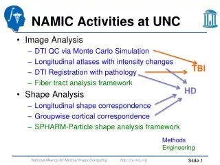
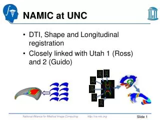
NAMIC Activities at UNC Methods Engineering • Image Analysis • DTI QC: error estimation via MC • DTI Registration with pathology • Longitudinal atlases with intensity changes • Fiber tract analysis framework: Atlas builder, DTIReg modules • Shape Analysis • Interactive surface correspondence • Longitudinal shape correspondence • Normal consistency in surface correspondence • Brain shape regression (application) • Validation • Human-like DTI/DWI software phantom • DTI tractography challenge MICCAI 2012
DTI QC • DTI/DWI noisy, artifact rich => QC needed • Correct motion & eddy currents • Reject bad gradients • How much rejection is still okay? • Simple threshold on numbers of rejected DWI? • Goal: Estimate errors in DTI wrt local direction • Use of Monte Carlo simulation at given SNR
Uniformity of Directions 30 dir Philips 42 dir 6 dir • Different sequences • No rejection • Same SNR ~ 10 • ΔFA: Average error in FA (SimFA = 0.4) • ΔPD: Average error in local orientation • Non-uniform Philips sequence worst ~50% higher orientation & ~75% higher FA error
Rejection Affects Uniformity Clustered rejection 25% larger error in orientation and 20% larger FA error Allows specification of threshold wrt Error
Validation: Tractography • Soft/hardware DTI phantoms not realistic • Goal: Create human brain like phantom • Inspiration: MNI-Brainweb • Use real data to create a synthetic phantom • Estimate fiber anatomy from real data • Estimate brain morphometry population • Sample/simulate brain morphometry • Apply morphometry to fiber anatomy • Compute DWI from simulated fiber anatomy • Evaluate tractography vs known ground truth
Fiber Anatomy • MICCAI 2012 workshop • Fiber Anatomy • 6 subjects at 1.5mm3 & 42 dir • High resolution atlas via unbiased group-wise registration • DWI atlas • Two-fiber tractography • Whole brain for overall anatomy • 20 GB of fibers… • Merge with individual tracts • Cerebro-spinal tract
Brain Shape Space • Training data: T1 UNC Normal Study • 100 subjects 20-60 • Brain Shape Space • Deformation fields to prior template • PCA over deformation fields • Gaussian generative sampling in PCA space • Find 3 closest training samples • Weighted unbiased atlas building • Weights relative to distance between training and generated sample • Simple, imperfect, but appear realistic
Validation/Evaluation • Simulate via CHARMED (Assaf/Basser) • Restricted diffusion within fibers (cylindrical) and hindered diffusion outside fibers (tensor) • Different Noise levels, DWI resolution, Gradient sampling scheme • Amazingly this was the hardest… • Evaluate geometry (Fillard/Gouttard) • CurveCompare tool • Dice overlap, probabilistic fiber-dice, error in position, tangent vector& curvature • Accuracy & Reliability
Next • Improve brain shape space • Explicitly model subcortical and cortical shape • Stats on inflated cortical surface via magnitude and orientation to original cortex • Partial nested sphere stats, PCA on principal components • Simulate pathology, tumors, TBI • Utah tumor simulator • How can we simulate TBI?
Other stuff… • Other NAMIC ongoing projects • DTI registration with pathology • Incorporating full brain tractography • Incorporating intracranial cavity landmarks • Brain shape regression for craniosynostosis • Cortex correspondence via SPHARM spherical registration of sulcal curves • MR Texture feature for disease appearance quantification
Pubs MICCAI: 2 conf & 3 workshop papers 8 SPIE submissions 2012 NAMIC journal papers: H. C. Hazlett, H. Gu, R. C. McKinstry, D. W. W. Shaw, K. N. Botteron, S. R. Dager, M. Styner, C. Vachet, G. Gerig, S. J. Paterson, R. T. Schultz, A. M. Estes, A. C. Evans, J. Piven, the IBIS Network, “Brain Volume Findings in 6-Month-Old Infants at High Familial Risk for Autism.,” Am J Psychiatry, vol. 169, no. 6, pp. 601–608, Jun. 2012. X. Geng, S. Gouttard, A. Sharma, H. Gu, M. Styner, W. Lin, G. Gerig, and J. H. Gilmore, “Quantitative Tract-Based White Matter Development from Birth to Age Two Years,” NeuroImage, pp. 1–44, Mar. 2012. O. Lindberg, M. Walterfang, J. C. L. Looi, N. Malykhin, P. Ostberg, B. Zandbelt, M. Styner, B. Paniagua, D. Velakoulis, E. Orndahl, and L.-O. Wahlund, “Hippocampal Shape Analysis in Alzheimer's Disease and Frontotemporal Lobar Degeneration Subtypes.,” J Alzheimers Dis, Mar. 2012. J. J. Wolff, H. Gu, G. Gerig, J. T. Elison, M. Styner, S. Gouttard, K. N. Botteron, S. R. Dager, G. Dawson, A. M. Estes, A. C. Evans, H. C. Hazlett, P. Kostopoulos, R. C. McKinstry, S. J. Paterson, R. T. Schultz, L. Zwaigenbaum, and J. Piven, “Differences in White Matter Fiber Tract Development Present From 6 to 24 Months in Infants With Autism.,” Am J Psychiatry, Feb. 2012. Y. Li, J. Gilmore, J. Wang, M. Styner, W. Lin, and H. Zhu, “TwinMARM: Two-stage Multiscale Adaptive Regression Methods for Twin Neuroimaging Data.,” IEEE Trans Med Imaging, Jan. 2012. Y. Shi, S. J. Short, R. C. Knickmeyer, J. Wang, C. L. Coe, M. Niethammer, J. H. Gilmore, H. ZHU, and M. Styner, “Diffusion Tensor Imaging-Based Characterization of Brain Neurodevelopment in Primates.,” Cerebral cortex (New York, N.Y. : 1991), Jan. 2012. E. Maltbie, K. Bhatt, B. Paniagua, R. G. Smith, M. M. Graves, M. W. Mosconi, S. Peterson, S. White, J. Blocher, M. El-Sayed, H. C. Hazlett, and M. Styner, “Asymmetric bias in user guided segmentations of brain structures.,” NeuroImage, vol. 59, no. 2, pp. 1315–1323, Jan. 2012. A. E. Lyall, S. Woolson, H. M. Wolfe, B. D. Goldman, J. S. Reznick, R. M. Hamer, W. Lin, M. Styner, G. Gerig, and J. H. Gilmore, “Prenatal isolated mild ventriculomegaly is associated with persistent ventricle enlargement at ages 1 and 2.,” Early Hum. Dev., Mar. 2012.
Principal Nested Spheres K sample points, N samples, vnk is the kth normal for the nth sample Main idea - Evaluate entropy across different objects for the kth correspondent normal Given v1k, …, vnk in unit sphere S2, fit a great circle δ(c) to minimize the sum of squared deviations of vnk from the great circle Find the Frechet mean on δ(c) PCA on S2=>Compute principal scores Normals projected into the unit sphere Great circle that approximates the data