Download
1 / 50
530 likes | 865 Vues
Chronic Myeloproliferative Neoplasies (CMPN). Myeloid Neoplasies WHO Classification. Myeloproliferative neoplasies (MPN) Myelodysplastic syndromes (MDS) Myelodysplastic / Myeloproliferative neoplasies Chronic myelomonocytic leukemia (CMML) Juvenile myelomonocytic leukemia (JMML)

E N D
MyeloidNeoplasiesWHO Classification • Myeloproliferativeneoplasies (MPN) • Myelodysplasticsyndromes (MDS) • Myelodysplastic /Myeloproliferativeneoplasies • Chronicmyelomonocyticleukemia (CMML) • Juvenilemyelomonocyticleukemia (JMML) • AtypicalChr.. MyeloidLeukemia (BCR/ABL -) • EosinophiliaandPDGFRA/PDGFRB orFGFR1mutation + myeloidorlymphoidneoplasies • Acutemyeloidneoplasies
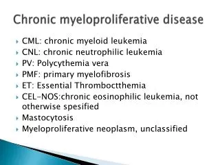
ChronicMyeloproliferativeNeoplasies (MPN) • Chronicmyelocyticleukemia (CML) (Bcr-abl +) • ChronicNeutrophylicLeukemia • PolycythemiaVera • PrimaryMyelofibrosis • EssentialThrombocythemia • ChronicEosinophylicLeukemia • Mastocytosis • Unclassifiedtypes
Systemicmastocytosis Polycythemia(rubra)vera Essentialthrombocytemia • Hypereosinophylicsyndrome Chronicmyeloidleukemia • Chronicmyelomonocyticleukemia Myelofibrosiswithmyeloidmetaplasia Myeloproliferative Neoplasies
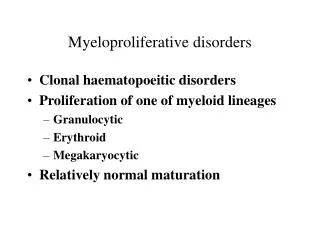
CMPN -concepts • Clonal disorders of hemopoietic stem cell • Overproduction of one or more cell lines • Extramedullary hematopoesis, myelofibrosis and leukemic transformation can occur • Increased risk of thrombosis
Polycythemia plasma Blood Cells (RBC) 6
1 2 3 a b DehydrationHct: elevated Hct: b/a Normal Truepolycythemia
Polycythemia • Relative • Dehydration • Gaisbock’s syndr (Nephropathy, Hypertension, Erythrocytosis) • Stress polycythemia • Absolute/true • Secondary • Primary • PRV • Familial
Erythropoetin Bone marrow Kidney Increasedredcellproduction Hypoksia Anemia Oxygen transport capacity of thebloodincreases IncreasedHblevel
Erythropoetin Bone marrow Kidney Increasedredcellproduction Causes of hypoksiaotherthananemia CO intoxication Highaltitude High O2 affinityHb Methemoglobinemia Lungdisease Respir. centerdysfunction Sleepapnea Righttoleftshunts Erythrocytosis (Polycythemia)
Increased EPO levelswithouthypoxia Bone marrow • High EPO duetoRenaldisease Renalcysts Hydronephrosis R.Arterystenosis Renaltransplantation Focalglomerulonephritis Bartter’ssyndrome Inappropriate EPO production • EPO productionbyTumors Hypernephroma Hepatoma Cerebellarhemangioblastoma Adrenal adenoma Pheocromocytoma Meningioma Uterinefibromyoma • Androgentherapy • EPO administration • Other Congenital /familialerythrocytosis Eg: EPO receptorhypersensitivity Increasedredcellproduction Erythrocytosis (Polycythemia)
Stemcelldisease Bone marrow Overproduction of RBCs + WBCs+ Platelets Polycythemiavera
PV (PRV) • FirstdescribedbyVaquez in 1892 • Epidemiology: • Incidence:2/100.000-year • Medianage: 60 (mayoccur in youngeralso, 7% < 40 y) • M/F: 1-2:1 • General characteristics: • Erytrocytosis,leukocytosis,thrombocytosis • Thrombosis, • Bleeding • Transformation: • Myelofibrosis/acuteleukemia
PRV: Etiology-Pathophysiology • PRV erythroid progenitors are resistant to apoptosis induced by EPO deprivation. • Non-EPO dependent overproduction of erythroid lineage • Increased sensitivity to cytokines or HGF : eg: IL-3, GM-CSF, SCF, TPO, IGF-1, • JAK-2 mutation !!!!!!!!!!!!!
PRV: Etiology-Pathophysiology • Progenitors in PRV gainclonalpredominanceandsupresstheproliferation of normal progenitorcells. • Duringthelaterstages of thediseasemyelofibrosisandextramedullaryhematopoesis (in theliver , spleenandlymphnodes) mayoccur.
PRV:Etiology-Pathophysiology • JAK2 mutations (chr 9p):TK activity: 97%* • Increased levels of PRV-1 mRNA in granulocytes *Kenneth Kaushansky, On the Molecular Origins of the Chronic Myeloproliferative Disorders: It All Makes Sense.ASH Education Book 2005,p 533-537
Campbell P and Green A. N Engl J Med 2006;355:2452-2466 JAK2 mutation (Chr 9p): 97% of cases !!!!!!! EPO unrelated signal EPO related signal
PRV: Clinical features • Asymptomatic • Plethora,cyanosis, • Vertigo • Tinnitus • Headache • Visualdisturbances • Paresthesias • Systolichypertension • Erythromelalgia • Pruritus (aggravatedbybath) • Weightloss • Arthralgia • Decreasedexercise • capacity, • Dyspnea • Excessivesweating • Vascularocclusion • TIA , stroke,MI • Venousthrombosis • Digitalischemia • Easybruising • GI bleeding • Splenomegaly (70%) • Goutor/andkidneystones • Pulmonaryhypertension
PRV: Clinical features • Late stage disease • Secondary bone marrow fibrosis and extramedullary hematopoesis • Increased spleen size and/or hepatomegaly • Decreasing red cell mass • Leuko-erythroblastic blood picture with tear drop poikilocytosis • Bone marrow fibrosis
Disease presentation: • Somecaseshaveisolatederythrocytosis • Somemayhavetrilinegaehyperplasia • erythrocytosis • leukocytosis • thrombocytosis • Somecasesmaypresentwithisolatedlekocytosisorthrombocytosis in thebeginning of thediseasefollowedbyerythrocytosis .
PRV • Hct > 60% in men and > 55% in women usually indicates red cell mass increase • Iron deficiency or hypersplenism may cause an underestimation of red cell mass due to a decreased Hct
Serum levels of • LDH • Uric acid • Vit B12 or B12 binding capacity may increase and • EPO is decreased • Leukocyte alkaline phosphatase level increases • JAK2 (+) • Red cell mass is increased • Bone marrow is • hypercellular (trilineage hyperplasia, red cell and megakaryocytic lineage may be prominent) and , • iron strores may be decreased. • fibrosis may be a late finding
Diagnosis • Excludeothercauses of polycythemia • Searchforthecriteriaforthediagnosis of PRV
PV 2008 WHO Diagnostic Criteria Major criteria Increased RBC mass Hgb>18.5 g/dL (male) >16.5 g/dL (female)or Hgb ya da Hct >99 percentile or Increased RBC mass (> % 25 of normal)(Cr51) JAK2V617F (%90-95) or JAK2 exon 12 mutation (%5-10) Minor criteria Bone Marrow: Proliferation of all three cell lines Serum EPO: Decreased Endogenous erythroid colonies • 2 major and 1 minör or • First major and 2 minor criteria are needed for diagnosis
Complications • Myelofibrosis • Acute leukemia • Peptic ulcer • Vascular occlusions:Arterial or venous • Bleeding:Platelet defects , acquired vWD • Splenic infarction • Increase in uric acid levels and gout or renal stones
PRV: Prognosis • Proliferative phase followed by postpolycythemic myelofibrosis and extramedullary hematopoesis • Life expectancy: > 10 years • Less than normal age/sex group • Thrombosis • Transformation • AML • MF
PRV Treatment • VenesectiontokeepHct: < %45 • Lowdose aspirin (ifit’s not contraindicated) • Prevent/controlreversiblethrombotic risk factors • EG:Hypertension,smoking, obesity, hypercholesterolemia,DM etc • Myelosupressivetreatment : Ifthere is; • Thrombosis • Age > 60 • Intolerancetophlebotomy • Prominentthrombocytosis • Symptomatic/progressivesplenomegaly
Myelosupressive agents: • Hydroxyurea • IFN • Busulphan • Radioactive “P” (32P) • Anegrelide (only to supress platelet production)
PV: TreatmentRisk group Treatment Venesection Low dose aspirin + Hydroksyurea (anegrelide or interpheron) High risk patients: • Thrombosis history+ • Age > 60 • Platelets > 1.500.000/mm3 Intermediate risk • Without high/low risk features Low risk: • Age < 40 • Without any risk factor Low dose aspirin + Venesection Myelosupressive treatment if CVS risk factors are present. Low dose aspirin + Venesection Peter J. Campbell and Anthony R. Green. Management of Polycythemia Vera andEssential Thrombocythemia,ASH Education Book 2005,p201-208
Causes of thrombocytosis • ET and other MPD • Iron deficiency anemia • Splenectomy or hyposplenism • Malignancy • Collagen vascular disease • Infection • Hemolysis or bleeding • Myelodysplastic Syndrome • Surgery • Drugs,etc
Essential Thrombocytemia(ET) • First described by Epstein and Goedel in 1934 as “haemorrhagic thrombocytemia” • In 1951 Dameshek defined it as a MPD
EssentialThrombocytemia (ET) • 1-2/100.000- year • Age: Median:50-60 • Second peak at age 30 (mostly female) • M/F:slight female predominance • Median survival > 10 years • Etiology : unknown • JAK2 mutation: 57 %
ET: Clinical • Some patients may be asymptomatic • Vasomotor symptoms: • Visual disturbances • Lightheadedness • Headaches • Palpitations • Atypical chest pain • Erythromelalgia • Livedo reticularis • Acral paresthesias
ET: Clinical • Thrombosis: • At presentationor • duringthecourse of thedisease • DeepVeinThrombosis, PulmonaryEmbolism • Digitalischemia • Portalveinthrombosis • CVA • Coronaryischemia • Bleeding: • Mostcommon : GIS • NSAID increasethe risk • Majorbleeding:5% • Splenomegaly : 20-25%
Diagnosis of ET: WHO 2008 4 criteria must be present Thrombocytosis > 450 x 10^9/L More than two months on more than two occasions Megakaryocytic proliferation without granulocytic and erythroid proliferation Without diagnostic criteria of CML,PV,IMF,MDS and other CMPN JAK2V617F (50%) or cMPL mutation (MPLW515L and MPLW515K) + or no evidence of reactive thrombocytosis
Exclude other causes of thrombocytosis Reactive Anemias Iron deficiency Hemolytic Acute blood loss Post splenectomy Inflammation Bone marrow findings Normo-Hypercellular with megakaryocytic hypeplasia Normal iron score Normal erythropoesis Without significant fibrosis Diagnosis of ET: WHO 2008 (2)
Prognosis • Myelofibrosis: (spent phase) • Acute leukemia: • Mortality: mostly because of thrombosis or bleeding
ET: Treatment • Prevent/Manage cardiovascular risk factors • Low dose aspirin • if there is no contra-indication • Myelosupressive treatment: • High risk patients • > 60 y, • prior thrombosis, • plt > 1500000/mm3 • Thrombopheresis • Rapid reduction of very high plt counts
ET: TreatmentRisk group Treatment Low dose aspirin + Hydroksyurea (anegrelide or interpheron) High risk patients: • Thrombosis history+ • Age > 60 • Platelets > 1.500.000/mm3 Intermediate risk • Age: 40-60 • Without high risk features Low risk: • Age < 40 • Without any risk factor Low dose aspirin + Myelosupressive treatment if CVS risk factors are present. Low dose aspirin + Reference: Peter J. Campbell and Anthony R. Green. Management of Polycythemia Vera andEssential Thrombocythemia,ASH Education Book 2005,p201-208
Causes of myelofibrosis Idiopathicmyelofibrosisand PRV ,ET, CML, MDS Lymphoma,M Myeloma A.Leukemia (ANLL-M7,ALL) Hairycellleukemia Metastaticcarcinoma Infection :eg: Tbc,Fungalinf. SLE,Systemicsclerosis SystemicMastocytosis Hypovit. D , renalosteodystrophy Hypoparathyroidism,Hyperparathyroidismetc
Etiology : Unknown (radiation ? other?)Epidemiology: 0.4 – 1.3 / 100000-yearAge: Median 60-65 (wide age distribution)Survival: median:3-5 years Myelofibrosiswithmyeloidmetaplasia (Idiopathicmyelofibrosis)
(Idiopathic Myelofibrosis) (Myelofibrosis with Myeloid Metaplasia) bone
IMF • Fibroblastsare not part of theclonalprocess • Megakaryocytederivedcytokines • PDGF • TGF-Beta arethecause of fibrosis • There is extramedullaryhematopoesis • Cytogeneticabnormalitiesarefound in 50% • 13 q- , 20q- , 12p- , trisomy 8, trisomy 9 • JAK2 mutation (50%) (orother . Eg MPL)
IMF:Clinical • Symptoms related to anemia or marrow failure • Hypermetabolic state • Sweating • Weight loss • Low grade fever • Oedema • Abdominal fullness/organomegaly (splenomegaly and or hepatomegaly) • Portal hypertension • Splenic infarction (sudden LUQ pain) • Effusions • LAP
IMF: LAB • Anemia • WBC : normal / leukopenia / leukocytosis • Plt : normal /thrombocytosis /thrombocytopenia • Blood smear : • Red cell poikilocytosis with teardrop shapes • Leuko-erythroblastic : erythroblasts + young myeloid cells* *(promyelocytes, myelocytes , meta , band and polys.)
IMF: LAB • LDH , uric acid: increased • Bone marrow : • Aspiration : Dry tap • Biopsy: Increased fibrosis and megakaryopoesis JAK2 mutation:50%** ** Kenneth Kaushansky, On the Molecular Origins of the Chronic Myeloproliferative Disorders: It All Makes Sense.ASH Education Book 2005,p 533-537
IMF: Diagnosis • Typicalbloodfindings • Splenomegaly • Bone marrow • Dry tap aspirate • Bone marrowfibrosisandmegakaryocyticdiysplasia (biopsyfinding) • JAK2 ( about 50%) • Othercausesexcluded
Complications • Transformationtoacuteleukemia • Gout • Infections • Terminationto a stage of deepmarrowfailure Prognosis • Deathdueto • Infection • ANLL (20% in first ten years)
IMF: Treatment • Transfusionswhenindicated • Androgens • Corticosteroids & other Myelosupressivetreatment • Hydroxyurea Other • Splenectomy • Splenicirradiation? StemCellTransplantation : • Theonlytreatmentwithcurepotential • Can be performed in a limitednumber of patients