Download
1 / 57
570 likes | 687 Vues
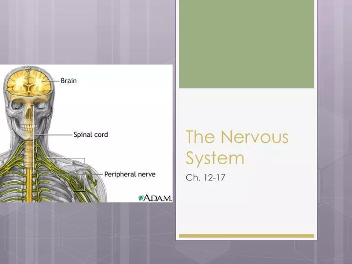
The Nervous System. Ch. 12-17. The Nervous System Introduction. Composed of Brain Spinal Cord Nerves 2 Major Subdivisions CNS (Central Nervous System) Brain, Spinal Cord PNS (Peripheral Nervous System) Cranial and Spinal Nerves 2 Major Subdivisions Afferent incoming pathways

E N D
The Nervous System Ch. 12-17
The Nervous System Introduction • Composed of • Brain • Spinal Cord • Nerves • 2 Major Subdivisions • CNS (Central Nervous System) • Brain, Spinal Cord • PNS (Peripheral Nervous System) • Cranial and Spinal Nerves • 2 Major Subdivisions • Afferent incoming pathways • Efferent Outgoing pathways
Neurons • Remember neuron structure: • The Cell Body or Soma • Contains a nucleus surrounded by cytoplasm along with all other organelles found within a cell • The Dendrites • Extensions from Cell Body/Soma • Receives information • The Axon • Single, long extension from Cell Body/Soma • Branch out into telodendria and end at synaptic/axon terminals
The Synapse • Each synaptic/axon terminal is part of a synapse • Site where the neuron communicates with another cell • Presynaptic Cell: sends the message (usually a neuron) • Postsynaptic Cell: receives the message (any cell type) • After a series of electrical signals is transmitted through the neuron, the axon/synaptic terminals release chemicals or neurotransmitters to the dendrite of another cell to continue the message
Synaptic Cleft Synapse
Neuron Video • http://www.youtube.com/watch?v=r71RoIkftd4
Neuroglia • Higher quantity and variation in CNS than PNS • Central Nervous System • Ependymal Cells • Astrocytes • Microglia • Oligodendrocytes • Peripheral Nervous System • Schwann Cells • Satellite Cells
CNS Neuroglia Ependymal Cells Microglia Removes cell debris, wastes, and pathogens by phagocytosis • Line ventricles in the brain and spinal cord • Aid in producing, circulating and monitoring of cerebrospinal fluid
CNS Neuroglia Astrocytes Oligodendrocytes Myelinate axons Provide structural framework • Provide structural support • Regulate ion, nutrient, and dissolved gas concentrations • Absorb and recycle neurotransmitters • Form scar tissue after injury
PNS Neuroglia Schwann Cells Satellite Cells Surround neuron cell bodies Regulate gas, nutrient, and neurontransmitter levels • Surround all axons • Responsible for myelination • Participate in repair process after injury
Synaptic Activity • Messages move from 1 location to another in the form of action potentials along neurons • Also known as nerve impulses • Messages move across the synapse • Can be electrical • Can be chemical (use of neurotransmitter)
Electrical Synapses • Presynaptic and postsynaptic cell membranes are locked together • Local electrical currents and directly transferred from cell to cell = • very rapid electrical impulse • Efficient action potential transfer from cell to cell
Chemical Synapses • Presynaptic and postsynaptic cell membranes are NOT locked together • Message not guaranteed to be transferred to the next cell dependent on the amount of neurotransmitters released • Different neurotransmitters: • Acetocholyne: found at neuromuscular junction • Norepinephrine: excitatory effect, adrenaline • Dopamine: Parkinson’s disease (rigidity of muscles) • Serotonin: attention, emotion, responsible for depression • GABA: reduce anxiety
Spinal Cord and Spinal Nerves Chapter 13
Spinal Cord Anatomy • Posterior/Anterior/Lateral Horn of Gray matter • Anterior Median Fissure • Posterior Median Sulcus • Spinal Nerve (Posterior/Anterior Roots) • Spinal Ganglion
Spinal Cord Anatomy • Adult – 18 inches in length • Only reaches down to L1-L2 • Spinal Meninges: specialized membranes that surround the spinal cord • Dura Mater • Arachnoid • Pia Mater • Bacterial or vial infections of the Meninges Meningitis
PiaMater Meninges • Meshwork of connective tissue • Dense area of vessels Dura Mater Arachnoid Filled with cerebrospinal fluid (SPF)—shock absorber Spinal tap • Tough, fibrous outermost layer • Collagen fibers • Covered with blood vessels and adipose tissue • Epidural Space
Nerve Plexuses • Complex interwoven network of nerves Plexus. • Cervical Plexus • C1-C5 (muscles of neck, thoracic cavity) • Brachial Plexus • C5-T1 (muscles of pectoral girdle and upper limb) • Lumbar Plexus • T12-L4 • Sacral Plexus • L4-S4
Refluxes Chapter 13
Reflexes • Rapid automatic responses to a specific stimuli made by a receptor • Preserve homeostasis • Rapid adjustments • The Reflex Arc: wiring of a single reflex • Beings at the receptor • Ends at peripheral effector (ex: muscle fiber)
Reflexes • Sensory Receptors • Proprioreceptors: provide info about body position and muscle control • Vestibular Receptors: provide a sense of equilibrium • Cutaneous Receptors: touch, pressure, heat and cold • Photoreceptors: respond to light • Chemoreceptors: respond to taste • Nocioreceptors: pain receptors
Reflexes • Arrival of a stimulus and activation of receptor (pain) • Activation of a Sensory Neuron (action potential along axons of neurons spinal cord) • Information Processing (neurotransmitter released and sensation related to brain) • Activation of Motor Neuron (axons carry action potential back towards the origin of pain) • Response of Peripheral Effector (release of neurotransmitter to skeletal muscle fiber contraction pulls hand away from pain) • http://www.youtube.com/watch?v=wLrhYzdbbpE
Reflexes • Receptor Density • More dense the receptors = more “sensitive” the area • Upon stimulation… • Phasic: receptors respond with an initial burst of action potentials and rapidly decrease, even though stimulus continues • Tonic: receptors firing at a constant rate as long as the stimulus is applied • Lab on reflexes
The Brain and Cranial Nerves Chapter 14
Human Brain • Contains 98% of neural tissue • Weighs about 3 lbs • Covered by a neural cortex (superficial layer of gray matter • Main Areas • Cerebrum • Cerebellum • Diencephalon • Brain Stem
Cerebrum • Largest portion of the brain • 2 hemispheres • Controls thoughts, sensations, intellect, memory, and complex movements • Lobes (correspond with cranial bones) • Frontal • Parietal • Occipital • Temporal
Cerebrum • Landmarks • Longitudinal fissure (separates the 2 hemispheres) • Central Sulcus (separates the frontal from parietal lobes) • Lateral Sulcus (separates the frontal from temporal) • Parieto-occipital sulcus (separates the parietal from the occipital) • White Matter • Dense region of axons
Cerebrum • Motor and Sensory Areas • Frontal Lobe = Voluntary control of skeletal muscles • Parietal Lobe = conscious perception of touch, pressure, vibration, pain, temperature, and taste • Occipital Lobe = conscious perception of visual stimuli • Temporal Lobe = conscious perception of auditory and olfactory stimuli, speech • All Lobes = Integration and processing of sensory data and motor activities
Cerebellum • 2nd Largest portion of the brain • 2 hemispheres • 2 Primary Functions • Adjusting Postural muscles of the body • Programming and fine-tuning movements
Other Major Regions of the Brain • Diencephalon: • Thalamus: relay and processing centers for sensory information • Hypothalamus: emotions, autonomic function, hormone production, body temp regulation
Other Major Regions of the Brain • Brain Stem :link between cerebrum and brain stem • Mesencephalon: aka “midbrain”; visual and auditory information • Pons: sleep & respiration • Medulla Oblongata: regulate heart rate, blood pressure, digestion
Ventricles of the Brain • Chambers filled with cerebrospinal fluid (CSF) • Lateral Ventricles (2 in each hemisphere of cerebrum) • Third Ventricle • Interventricular Foramen (connection between the lateral ventricles and third ventricle) • Cerebral Aqueduct • (connection between the third ventricle and fourth ventricle) • Fourth Ventricle • Narrows and opens into the spinal cord
Protection and Support • Protected by • Bones of cranium: mechanical protection (car) • Cranial Meninges: anchor (seat belt) • Dura Mater: outer layer • Arachnoid: middle layer • Pia Mater: surface of brain • Cerebrospinal Fluid: cushions against shocks/jolts (air bag)
Cerebrospinal Fluid (CSF) • Function: • Cushion • Support • Transport nutrients/chemical messengers/waste products • Formation: • Choroid plexus (in all ventricles): specialized cells that produce CSF
Blood Supply to the Brain • Brain requires a tremendous amount of blood • Receives 15-20% of blood pumped by heart • Interruption unconsciousness/irreversible brain damage • Dependent on constant supply of oxygen & glucose • Receives blood through arteries
Hydrocephalus • Condition with infants • Prior to fusion of cranial bones • Excess CSF (due to blockage or constriction of the meninges)causes the skull to enlarge • Infants suffer some degree of mental retardation
Cranial Nerves • Part of the PNS • Connected to the brain and branch out • 12 of them • Attaches the brain near a sensory or motor neuron • 2 of each
Cranial Nerves • Olfactory Nerves smell • Optic Nerves Vision • Oculomotor Nerves Eye Movements • Trochlear Nerves Eye movements • Trigeminal Nerves Sensory/Motor to face • Abducens Nerves Motor eye movements • Facial Nerves Sensory/Motor to face • Vestibulocochlear Nerves balance/equilibrium and hearing • Glossopharyngeal Nerves Sensory/Motor to head and neck • Vagus Nerves Sensory/Motor to thorax and abdomen • Accessory Nerves Motor to muscles of neck and upper back • Hypoglossal Nerves Motor for tongue movements
“Oh, once one takes the anatomy final, very good vacations are heavenly”
Conscious/Unconscious • Conscious = state of awareness of external stimuli • Unconscious = number of conditions, deep, unresponsive state drifting into sleep
Levels of Sleep • 2 levels, patterns of brain activity • Slow wave sleep • Deep sleep/non-REM sleep • Entire body relaxed • Cerebral cortex activity at minimum • Heart rate, blood pressure, respiratory rate, energy utilization decline by 30% • Rapid Eye Movement (REM sleep) • Active dreaming • Changes in blood pressure/respiratory rate • Muscle tone decreases • Neurons controlling eye muscles stop regulation = eyes move rapidly as dream events unfold
Sleeping Cycles • REM and Deep sleep alternate throughout the night • Starts with Deep sleep = 1.5 hours • REM last on average 5 minutes • Determined by an EEG • Electroencephalogram: graphic record of the electrical activities of the brain
Alzheimer’s Disease • Loss of cerebral functions • Symptoms appear around 50-60 years old • Can affect younger individuals but is rare • Currently affects 2 million ppl in USA • 100,000 deaths each year • Chromosome 14, 19, and 21 • Majority of Downs Syndrome develop it (21)