Download
1 / 72
730 likes | 756 Vues
CT lung. Technique. Depends on the indication Routine protocol High resolution protocol. Positioning Supine arms up Scan length From the base of the neck to The diaphragm The adrenals in cancer patients. Breathing Full inspiration Instruction

E N D
Technique • Depends on the indication • Routine protocol • High resolution protocol
Positioning • Supine arms up • Scan length • From the base of the neck to • The diaphragm • The adrenals in cancer patients
Breathing • Full inspiration • Instruction • Make sure there is adequate understanding • Rehearse • 4-5 deep breaths before start • 3-4 secbefore scan • Scan outwards
IV CM (routine) • 90 mls • 2ml/sec • 25 (20-30) secs scan delay • IV CM angiography • 90-110 mls CM + 40-50 mlsnormal saline • 3ml/sec • 20sec delay orbolus tracking • Oral prep • oesophagus • Tumor invasion • Just before scan
Section thickness • Thin originals • Thicker for viewing • Filters (raw data) • Standard (lungs mediastinum) • Bone HR lung parenhyma • Optical filters • WW/L • lungs: 1500 / -600 • mediastinum: 400 / 50
Exposure factors • 120kV • 150 mAs • CTDIvol5 - 10mGy • Due to high intrinsic contrast we can afford lower SNR and use lower exposure factors • The beam starvation artifacts in the shoulders and abdomen may be reduced by dose modulation techniques.
High Resolution CT (HRCT) • Thin sections < 2mms • Bone filter • Deep inspiration • Expiration - End expiration • Air trapping • DD air trapping – ground glass • Wall invasion by Cancer • Prone - inspiration • Only the suspicious areas usually lung bases • 5 mins before scan • Avoid the need by scanning immediately after the patient lies flat • ΜΙΡ - minIPin different levels
CTlow dose • Repeat scan for lung parenchyma assessment • DoseCTDIvol = 2-6mGy • Less sharp algorithm to manage noise • Higher kV (140) • Noise filtration
CT low dose – lung cancer screening • Thin sections (2-3mm) • Thicker reprocessing • No IV CM • CAD
Virtual bronchoscopy • 3D technique • No measurements
Congenital diseases • atresia http://radiographics.rsna.org/content/29/5/1531 και 1497
Agenesis, hypoplasia http://www.ajronline.org/content/183/5/1497
tracheobronchomegaly http://www.learningradiology.com/archives06/COW%20230-Mounier-Kuhn/mounierkuhncorrect.html
Bronchogenic cyst • Lung sequestration http://radiology.rsna.org/content/217/2/441.long http://emedicine.medscape.com/article/412554-overview#a20
Scimitar syndrome http://radiographics.rsna.org/content/27/5/1323.full http://www.ajronline.org/content/183/5/1497
Diseases of the airways • Stenosis tracheal • Use WL=-700 και WW>1000 • Movement may create double wall http://radiographics.rsna.org/content/22/suppl_1/S215
Bronchiectasis http://www.learningradiology.com/archives2011/COW 468-Cystic Bronchiectasis http://radiology.casereports.net/index.php/rcr/article/viewArticle/137/383
Bronchiolitis • Aspergillosis http://www.ajronline.org/content/185/2/354/F15 http://radiology.rsna.org/content/222/3/771
Tumors • pappiloma – • pappilomatosisΘηλωμάτωση • carcinoid http://www.ncbi.nlm.nih.gov/pmc/articles/PMC2627258/
Bronchial carcinoma http://jco.ascopubs.org/content/25/34/5521.full
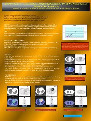
Lung nodules • Granuloma • Benign tumors • Hamartoma, chondroma http://emedicine.medscape.com/article/356271
Malignant tumors • Bronchogenic, alveolar cell, metastatic, carcinoid, kaposi sarcoma http://www.radiologyassistant.nl/en/42459cff38f02
Lung nodule • Benign vs malignant http://www.radiologyassistant.nl/en/460f9fcd50637
Pancoast tumor http://www.radiologyassistant.nl/en/42459cff38f02
Pancoast tumor http://www.radiologyassistant.nl/en/42459cff38f02
Staging – ΤΝΜ – Τ http://www.radiologyassistant.nl/en/42459cff38f02
StagingΤΝΜ – Ν http://www.radiologyassistant.nl/en/42459cff38f02
StagingΤΝΜ - Μ • Brain • Liver • Adrenals
Embolism http://imaging.consult.com/image/case
AV malformations http://www.vcuthoracicimaging.com/Historyanswer.aspx?qid=71&fid=1
http://www.vcuthoracicimaging.com/Historyanswer.aspx?qid=71&fid=1http://www.vcuthoracicimaging.com/Historyanswer.aspx?qid=71&fid=1
http://www.vcuthoracicimaging.com/Historyanswer.aspx?qid=71&fid=1http://www.vcuthoracicimaging.com/Historyanswer.aspx?qid=71&fid=1
Intrapulmonary lymphnodes • Round atelectasis http://www.learningradiology.com/notes/chestnotes/roundatelectasispage.htm
Infection • Bacterial pneumonia • Atypical pneymonia • Viral pneumonia • Tuberculosis • Pneumonia in the immunocompromised patient • Bacterial • Viral and pneumocystisCarinii, opportunistic or fungal
http://www.springerlink.com/content/hjnk528djcwwe8et • Atypical - viral
PneumocystisCarinii Pneumonia (PCP) immunocompromised http://www.springerlink.com/content/hjnk528djcwwe8et
Tuberculosis • TB – primary • consolidation • lymphadenopathy • milliary • effusion http://radiographics.rsna.org/content/27/5/1255/F4
TB - secondary • consolidation • cavities • fibrosis • bronchiectasis • empyema http://radiographics.rsna.org/content/27/5/1255/F4