Download

1 / 12
140 likes | 520 Vues
Salivary Gland Diseases. The salivary glands consist of 3 paired major glands, 1- parotid glands: opens against the upper 2 nd molar buccally by Stensen’s duct, the secretion is mainly serous.

E N D
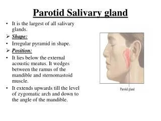
The salivary glands consist of 3 paired major glands, • 1- parotid glands: opens against the upper 2nd molar buccally by Stensen’s duct, the secretion is mainly serous. • 2- submandibular glands: opens near the lingual frenum by Warthin’s duct, the secretion is mixed but mainly serous. • 3- sublingual glands: open near the opening of submandibular gland by Bartholin’s duct, the secretion is mixed but mainly mucous. • In addition to these major glands, there is a countless of minor salivary glands found in almost every part of the oral cavity, except the gingiva & anterior region of the hard palate. • Both, major & minor salivary glands consist of parenchyma elements which are supported by C.T. stroma. • The paranchymal is derived from the oral epith & consist of terminal secretory units leading to ducts that open into the oral cavity. The parenchyma surrounded by a C.T. capsule & extend into it. • The blood & lymphatic vessels & nerves that supply the gland will contained within the C.T. • The normal function & health of the mouth depends on the normal composition & secretion of the saliva. • The important function of salivary glands is the production of saliva which contain various organic & inorganic substances & help in mastication, deglutition & digestion of food.
Investigations for salivary glands: • 1- Sialometery: measures the amount of saliva production in a certain time. • 2- Sialochemistry: measures the composition of saliva. • 3- Sialography: by introducing the iodine containing contrast media through the opening of the duct. • 4- Sonagraphy: Ultrasonic patterns when dealing with minor salivary glands. • 5- Cytology: by aspiration. • 6- Biopsy. • Classification of salivary glands diseases: • 1- Obstructions: this could be by calculi or cystic type (stone, mucocele) • 2- Infections: viral (Mumps), bacterial (acute & chronic Sialadenitis) • 3- Degenerative changes: Sjogren syndrome, radiation. • 4- Functional disorders. • 5- Neoplasms.
1- Obstructions:duct obstruction may resultfrom either:A- blockage of the lumen (calculi, mucocele)B- disease in or around the duct wall (fibrosis, neoplasia) • A- Sialoliths (S.G. stone): • Mean presence of calculi or stones within the duct. • The calculi believed to arise from the deposition of ca ++ salt around a nidus of debris within the duct lumen, these debris include bacteria, ductal epith cells, or foreign bodies. • 70-90% of stones occur in the submandibular gland, & this due to long tortuous path of the duct & thick secretion of the gland. about 6% in parotid gland & 2% in sublingual gland & minor S.G. • Mainly occur in adult male & is usually unilateral. • Symptoms: pain, sudden enlargement specially at meal time. • Radiography: there will be radiopaque mass, however, about 40% of parotid & 20% of submandibular stones are not radiopaque, therefore Sialography may be needed to locate them. • Treatment: • removing the calculi by manipulation or incision of the duct.
A common lesion of the oral mucosa it is of 2 types: 1- Mucus extravasation cyst: Result from rupture of a S.G. duct & spillage of mucin into the surrounding soft tissue, as a result of local trauma. Clinically,appear as a bluish or translucent swelling, soft, fluctuant, range from mms to cms. Mostly in child & adult. The lower lip is the most common site usually lateral to the midline. The duration of the lesion can vary from a few days to several years & many patients relate a history of a recurrent swelling that may periodically rupture & release it’s fluid contents. Mucus extravasation cyst is not true cyst, because it lacks an epith lining. Histopathology: An area of spilled mucin surrounded by a granulation tissue response. The inflammation includes numerous neutrophils & foamy macrophages. In some cases, a ruptured salivary duct may be identified feeding into the area. Treatment: surgical excision. B- Mucocele
2- Mucus retention cyst: • This derived from cystic dilatation of a duct, due to partial or complete obstruction of the duct, that make the mucin to remain (retention) within the duct. • Clinically, like the extravasation type. • Histopathology: • Cyst lining is variable (ductal epith in origin) composed of cuboidal, columnar or squamous epith, surrounding the mucoid secretion in the lumen. • Treatment: • Surgical excision.
3- Ranula • it is a type of extravasation mucocele, the source of mucin spillage is usually the sublingual gland or from submandibular duct or possibly from minor S.G. in the floor of the mouth. • Clinically, appear as swelling in the floor of the mouth resemble a Frog’s belly. • It may interfere with the speech or mastication, because it causes pushing of the tongue up toward the palate. • Treatment: • By total or partial removal or marsupulization.
2- Infections • A- Viral infection (Mumps): • Is an acute, contagious infection which often occurs in minor epidemics & is caused by Paramyxovirus. • It is the commonest cause of parotid enlargement & may affect the submandibular & sublingual glands. • The virus transmitted by direct contact with infected saliva & by droplet spread. Mostly affect the children & the incubation period is about 2-3 weeks. • Clinically, the disease start with fever, malaise, followed by painful swelling of sudden onset behind the ear. • The bilateral parotid involvement occur in about 70%. • Then the swelling gradually subsides over a period of about 7 days. • Occasionally, in adults other internal organs are involved, such as testes, ovaries, CNS, & pancreas. Orchitis is the most common complication, occurring in about 20% in adult males. • After the attack, immunity is long-standing, & with use of vaccine, childhood mumps becomes infrequent. • B- Pyogenic bacterial infections:are common & may be seen after major abdominal surgery or in glands that have been obstructed.
3- Degenerative disease • Sjogren Syndrome • Is an immune-mediated chronic inflammatory disease, characterized by lymphocytic infiltration & acinar destruction of salivary & lacrimal glands. • Mainly affects middle-aged females, & symptoms related to dryness & soreness of the mouth & eyes are common clinical presentations. • The patient also complain from difficulty in swallowing & speaking, increased fluid intake, disturbance of taste, & rapidly progressive caries. • S.G. enlargement is usually bilateral without pain, & predominantly affects the parotid gland. • The disease classified into 2 types: • 1- primary: xerostomia + xerophthalmia • 2- secondary: xerostomia + xerophthalmia + C.T. disease usually rheumatoid arthritis. • Histopathology: • Initially, the S.G. show lymphocytic infiltration around intralobular ducts with acinar atrophy & obliteration of the duct lumen by proliferation of ductal epith, lead to formation of islands of epith tissue, termed epimyoepithelial islands. • Finally, the lesion consists of sheets of lymphoid cells surrounding the epimyoepithelial island & replacing entire S.G. lobules.