Download
1 / 23
240 likes | 429 Vues
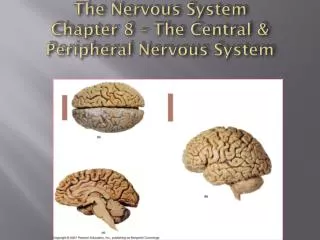
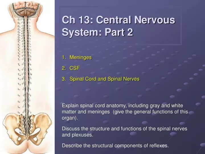
Ch 13: Central Nervous System: Part 2. Meninges CSF Spinal Cord and Spinal Nerves. Explain spinal cord anatomy, including gray and white matter and meninges (give the general functions of this organ). Discuss the structure and functions of the spinal nerves and plexuses.

E N D
Ch 13: Central Nervous System: Part 2 Meninges CSF Spinal Cord and Spinal Nerves Explain spinal cord anatomy, including gray and white matter and meninges (give the general functions of this organ). Discuss the structure and functions of the spinal nerves and plexuses. Describe the structural components of reflexes.
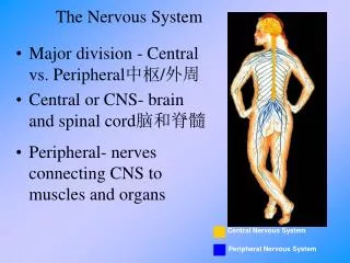
1. Cranial Meninges Three layers: 1. Dura mater - strong, "tough mother" a. falxcerebri b. falxcerebellic. tentoriumcerebelli 2. Arachnoid - spidery, holds blood vessels 3. Pia mater - "delicate mother" Note: Subdural hematoma
Formation in ventricles by specialized ependymal cells of choroid plexus(~500 mL/day; total volume ~ 150 mL) Functions transport medium (nutrients, waste) shock absorption buoyancy(floats the brain) CSF circulation: Ventricles → central canal → subarachnoid space An important diagnostic tool. 2. CSF: Cerebrospinal Fluid
Longitudinal fissure Arachnoid granulations: This is where the CSF produced in the choroid plexuses of the ventricles and which has circulated into the subarachnoid space is reabsorbed.
Meningitis: inflammation of meninges/CSF • Bacterial • Relatively rare • Life threatening • Antibiotics • Fungal • Viral—most common • Younger • Self-resolving
Blood Brain Barrier (BBB) • Tight Junctions in capillary endothelium prevent passive diffusion into the brain. Lots of Active Transport, especially of H2O soluble compounds (think glucose). • Fat soluble compounds readily pass the BBB • E.g. steroid hormones, ADEK • Major role of astrocytes • 3 areas in brain don’t have BBB • portion of hypothalamus • pineal gland (in diencephalon) • choroid plexus
3. Spinal cord: • Resides inside vertebral canal • Extends to L1/ L2 • 31 segments, each associated with a pair of dorsal root ganglia • Two enlargements • Cervical and Lumbar • Conusmedullaris • CaudaEquina • FilumTerminale Fig. 13-29
Cervical Enlargement Gray matter expanded to incorporate more sensory input from limbs and more cell bodies for motor control of limbs
Lumbar Enlargement See fig 14-1
Spinal Meninges Three membranes surround all of CNS 1) Dura mater - "tough mother", strong. Note the Epidural Space. 2) Arachnoid - spidery looking, carries blood vessels, etc. Note the Subarachnoid space which contains CSF 3) Pia mater - "delicate mother", adheres tightly to surface of spinal cord 2a) Subarachnoid Space 3) Pia mater 2) Arachnoid 1) Dura mater
Transverse Section Fig 13.30 Compare the spinal roots with the model of the vertebral column in the lab. Note that the dura covers both the dorsal and ventral roots.
Organization of Spinal Cord Gray matter - interior horns posterior - somatic and visceral sensory nuclei anterior (and lateral) gray horns – somatic and visceral motor control gray commissures - axons carrying information from side to side White matter - tracts or columns posterior white column - anterior white column lateral white column anterior white commissure functions ascending tracts - sensory toward brain descending tracts - motor from brain
Sectional anatomy of spinal cord Outer white part; inner gray butterfly
Lumbar Puncture (= Spinal Tap) L3 S1 For clinical examination of CSF or administration of radiopaque dyes, drugs and anesthetics However: mostly “epidurals” for anesthetics
Organization of Spinal Nerves: 1. Root – inside vertebral canal a. dorsal sensory root with a ganglion b. ventral motor root 2. Mixed spinal nerve 3. Rami a. dorsal - mixed to skin and muscles of back b. ventral - mixed “spinal nerve” to ventrolateral body surfaces and limbs c. white ramus communicans motor ANS d. gray ramus communicans motor ANS
Reflexes Fast, preprogrammed, inborn, automatic responses Occur in the CNS at the spinal cord or brainstem levels (cranial nerves) May be either monosynaptic or polysynaptic All require a. stimulus at receptor b. sensory information relay c. processing at CNS level d. activation of motor response e. response of peripheral effector
Dermatomes • Sensory innervations by specific spinal nerves Each pair of spinal nerves monitors specific region of body surface.