Download
1 / 15
390 likes | 1.18k Vues
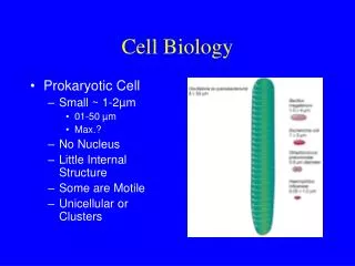
History of Cell Biology. Chapter 4, Section 1 Video: Brief History of Cells. Robert Hooke. English scientist (1635) Used a light microscope to study nature Looked at a thin slice of cork from the bark of a cork oak tree “great many little boxes”

E N D
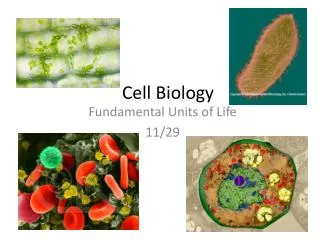
History of Cell Biology Chapter 4, Section 1 Video: Brief History of Cells
Robert Hooke • English scientist (1635) • Used a light microscope to study nature • Looked at a thin slice of cork from the bark of a cork oak tree • “great many little boxes” • Reminded him of cubicles or “cells” where monks lived
Anton van Leeuwenhoek • Dutch trader (1632) • 1st to observe living cells • Algae: Spirogyra • Protists: Vorticella • Animalcules (protists) • Made microscope 10x that which Hooke used • Known to have made over 500 different microscopes
Matthias Schleiden • German Botanist (1804) • Concluded that all plants were composed of cells
Theodor Schwann • German zoologist (1812) • Friend of Schleiden • Concluded that all animals are composed of cells
Rudolf Virchow • 1821 German Physician • Concluded that all cells come from other cells
Cell Theory • All living organisms are composed of one or more cells. • Cells are the basic units of structure and function in an organism. • Cells come from the reproduction of existing cells.
Compound Light Microscope • Two lenses to magnify an image • Eyepiece (ocular lens): magnifies usually 10x • Objective lens: enlarges the image • 4x, 10x, 40x • Stage: platform that supports the slide • Light Source: light bulb • Either light reflected with a mirror or an incandescent small lamp http://www.semsupplies.com/Resources/Image93.gif
Paramecium Elodea - green algae http://science.exeter.edu/jekstrom/JPEG/ELODEA~1.JPE • Specimen has to be thin enough • for light to pass through • Can see living organisms. http://ebiomedia.com/gall/classics/Paramecium/images/para8.jpg
Transmission Electron Microscope (TEM) • Transmits a beam of e- through thinly sliced specimen • Magnetic lenses enlarge the image • Can magnify up to 200,000x • Black and white • Can not view living specimens http://mstd.llnl.gov/highlights/matchar/images/tem.jpg
VENTRICULAR MUSCLE CELLS Virus in a fish gill cell http://www.pbrc.hawaii.edu/bemf/taura4_med.jpg http://www.ualberta.ca/~mingchen/pics/s-muscle.jpg
Scanning Electron Microscope (SEM) • Sprayed with a fine metal coating • Beam of e- over the specimen’s surface • Causes metal coating to emit a shower of e- • Project onto a fluorescent screen • Three-dimensional • Add color • Magnify up to 100,000x http://www.lbp.police.uk/forensicscience/images/scanning%20electron%20microscope.jpg
Pfiesteria-toxic dinoflagellates Red Blood Cells http://www.abc.net.au/science/slab/cells/img/cell.jpg http://www.physiol.usyd.edu.au/daved/teaching/images/rbc.jpg
Scanning Tunneling Microscope • Uses needle-like probe to measure differences in voltage caused by e- • Computer tracks the movement of the electrons • Gives 3D image • Adds color http://www.engr.unl.edu/erc/images/afm.jpg
Gold Nanoparticles http://www.chem.utoronto.ca/staff/DHIRANI/stm-np.jpg