Download
1 / 29
290 likes | 398 Vues
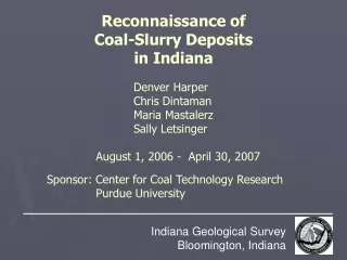
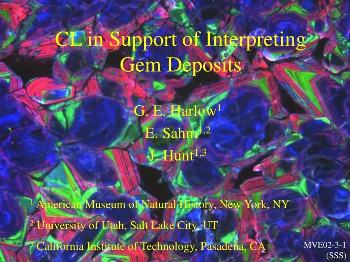
CL in Support of Interpreting Gem Deposits. G. E. Harlow 1 E. Sahm 1,2 J. Hunt 1,3. 1 American Museum of Natural History, New York, NY 2 University of Utah, Salt Lake City, UT 3 California Institute of Technology, Pasadena, CA. MVE02-3-1 (SSS). Instrumentation.

E N D
CL in Support of Interpreting Gem Deposits G. E. Harlow1 E. Sahm1,2 J. Hunt1,3 1 American Museum of Natural History, New York, NY 2 University of Utah, Salt Lake City, UT 3California Institute of Technology, Pasadena, CA MVE02-3-1 (SSS)
Instrumentation • Hitachi S-4700 Field-Emission SEM with BSE, EDS and a Gatan MonoCL3 detector & monochromator system (Peltier-cooled high-sensitivity PMT). The grating is a low-dispersion (21.6nm / mm slit width), with peak response (blaze wavelength) at 500nm and useful range of ~200 - 1200 nm.
CL Applications • Cathodoluminescence is an extremely powerful technique for examining zoning in minerals and can lead to fundamental interpretations about how they formed. • Targets in this presentation: • Jadeitites: jadeite, zircon, grossular? • Corundum deposits: ruby, sapphire, painite
Guatemalan Jadeitites • Jadeitites have been interpreted as crystallizations from aqueous fluids derived from subductions channels based, in large part, on CL observations. • In Guatemala there are two distinct serpentinite mélanges containing jadeitite, North and South of the Motagua fault. • How do the CL signatures of the minerals compare between the two distinct sources.
Phengite Qtz Jd-2 Zrn Omph Jd-1 Ttn SOUTH MVE02-8-6 Phengite Jadeitite, San Jose
Panchromatic Jadeite-phengite rock (MVE02-8-6 – SOUTH) Jadeite 480 nm Blue 270 nm UV Jd-2 Jd-1 SOUTH
Zircon, Secondary Electrons Jd-2 Zrn Jd-1 BSE Zircon Panchromatic Jadeite-phengite rock(MVE02-8-6 –SOUTH) Zircon 500 315 240 410 Panchromatic
315 nm UV 410 nm Blue 505 nm Green 685 nm Red
Zircon in lawsonite-eclogite(MVE02-6-1 – SOUTH) Zircon, Panchromatic 490 230 230 nm UV 490 nm Blue-green
Pmp Grs Jd BSE Pmp-Grs-Jadeitite(MVE04-20-1 – SOUTH) Jadeite 330 600 940 Panchromatic CL
MVE04-20-1 Pmp-Grs-Jadeitite Jadeite SOUTH BSE Panchromatic CL Panchromatic CL
MVE04-20-1 Pmp-Grs-Jadeitite Jadeite 300 nm UV Panchromatic 600 nm Green 940 nm IR
Pmp Grs Jd BSE Panchromatic Pmp-Grs-Jadeitite(MVE04-20-1 – SOUTH) Grossular 590 335 ~850 475 Panchromatic
NORTH Jadeitite, Río La Palmilla, North of MFZ(MVJ84-9D) 2 cm across (courtesy of S. Sorensen)
Panchromatic CL image of same area. Note healed fracture in jadeite grain. NORTH
Altered Jadeite (MVJ84-9B)at boundary between Jd and Ab+Ne NORTH
Room Temperature Spectrum Imaging Scale bar is 1 about micrometer. Spectrum Image (40 X 40 pixels) of adjacent area NORTH
Point 0 Point 1 Nepheline? Albite? Darker Jadeite Lighter Jadeite ~700 ~560 ~700 ~560 ~480 Point 2 Point 3 Room temperature CCD CL spectracourtesy of Paul Mainwaring
Zircon, Meta(?)-Jadeitite(MVJ84-9C – NORTH) 547 279 350 279 nm UV 350 nm UV 547 nm Green
Jadeite-Phengite rock (MVE02-8-6 -- SOUTH) • Comparison of CL Spectra from Jadeite • UV to IR peaks, but too soon to make generalizations other than color zoning observed by Sorensen and co-workers. 480 270 Pmp-Grs-Jadeitite(MVE04-20-1 -- SOUTH) Altered Jadeite(MVJ84-9B -- NORTH) Jadeite 330 ~700 ~560 ~480 600 940
Zircon in lawsonite-eclogite (MVE02-6-1 -- South) • Comparison of CL spectra from zircon • Main peak near 500nm and lowest near 250nm, but … • All are features show only primary growth. 490 230 Jadeite-phengite rock(MVE02-8-6 -- South) Zircon, Meta(?)-Jadeitite(MVJ84-9C -- North) Zircon 547 500 315 240 279 350 410
BSE Ruby (107643), Mogok, Burma inMarble w/ Spinel, Blue Cancrinite, Sodalite, Scapolite, Phlogopite, etc. 694nm - Cr 320 840 Panchromatic
320 nm UV 695 nm Cr-ruby Artifact of mirror 840 nm IR 840 nm IR
Ruby-1, Namya, CL Ruby-1, Namya, BSE Ruby-3, Namya, BSE Ruby-3, Namya, CL
Painite (CaZrBAl9O18) from Namya, Myanmar – rare gem mineral probably grew during skarn formation CL shows fine growth layering, implying growth pulses. Inclusions: Cc, Baddeleyite (ZrO2) & Srilankaite (TiZr2O6) Panchromatic
Some Conclusions • CL in zircon, jadeite and garnet of jadeitites is likely due to REE based on enrichment in these rocks. • Zoning structures suggest growth from fluids – for Zircon in jadeitite this implies growth at T = ~ 300°C at P > 1 GPa. • Considerable spectral structure from UV to IR is seen via SEM/CL. • Lots more to do.
Many Thanks to: • Jade & Ruby Helpers: Jinny Sisson, Sorena Sorensen, Carlos Gonzales, Mauricio Chiquin, Will Maze, Bill Larson, George Rossman, Jamie Newman, U Han Htun, Dr. Saw Naung U, Mint Soe • The Founders of the Feast: AMNH, NMNH, Frohlich Charitable Trust, Astor Expedition Fund,Sprague Fund, Michael Scott, & the National Science Foundation.