Download
1 / 71
740 likes | 1.15k Vues
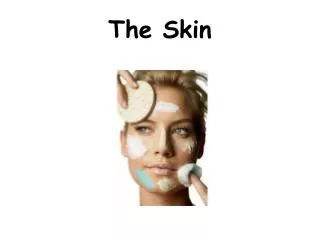
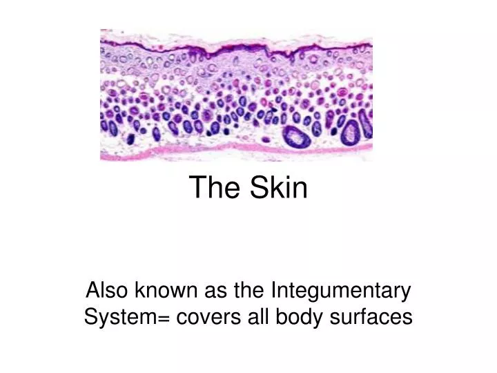
The Skin. Also known as the Integumentary System= covers all body surfaces. Skin Video. Contains these tissues:. 1. Epithelial Tissue a. Found in outer layer of skin (covers body) 2. Connective Tissue a. Consists of tough, flexible fibers that holds the body together. 3. Muscle Tissue

E N D
The Skin Also known as the Integumentary System= covers all body surfaces
Contains these tissues: • 1. Epithelial Tissue • a. Found in outer layer of skin (covers body) • 2. Connective Tissue • a. Consists of tough, flexible fibers that holds the body together. • 3. Muscle Tissue • a. Push hairs up in skin (goose bumps) • 4. Nervous Tissue • a. Helps detect pain, pressure, heat, cold
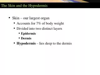
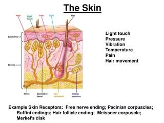
Consists of three major regions • Epidermis – outermost superficial region • Dermis – middle region • Hypodermis (superficial fascia) – deepest region
Functions • Covers all body surface: • inside and out • Ex. Lung linings • capillaries • anchored by basement membrane • reproduce rapidly • tightly packed • attached by desmosomes
The Two Layers of the Skin(Outer and Inner) • 1. Epidermis • a. Outermost layer of skin • 1. Exterior • Consists of 25 to 30 layers of dead cells (continually being shed) • Contains keratininized stratified squamous epithelium - protective protein • Contains keratinocytes, melanocytes, Merkel cells, and Langerhans’ cells
b. Characteristics • 1. No blood vessels • 2. Newer cells below push up to surface • 3. Older cells layer up and die • 4. Hardening = keratinizing • 5. calluses form • 6. shields against moisture loss • 7. Prevents invasions of organisms • 8. Ability to tan
Over view…. • 2. Outer portion of the skin is exposed to the external environment and functions in protection • 3. Interior • Contains living cells (continually forming new cells) • Melanin (pigment) found in this layer
3. Epidermal ridges • a. Known as fingerprints
Types of Fingerprints: • 1. Arch
Layers of Epidermis • From outer to inner: • 1. Stratum Lucidum = (Clear Layer) • 2. Stratum Granulosum = (Granular Layer) • a. Granular in appearance 3. Stratum Spinosum = (Prickly Layer) • a. Thick part • 4. Stratum Basale = (Basal Layer)b a. Deepest layer 5. Stratum Corneum = (Horny Layer) a. Layer of hard cells
Layers of the Epidermis: Stratum Corneum (Horny Layer) • Outermost layer of keratinized cells • Accounts for three quarters of the epidermal thickness • Functions include: • Waterproofing • Protection from abrasion and penetration • Rendering the body relatively insensitive to biological, chemical, and physical assaults
Layers of the Epidermis: Stratum Lucidum (Clear Layer) • Thin, transparent band superficial to the stratum granulosum • Consists of a few rows of flat, dead keratinocytes • Present only in thick skin
Layers of the Epidermis: Stratum Granulosum (Granular Layer) • Thin; three to five cell layers in which drastic changes in keratinocyte appearance occurs
Layers of the Epidermis: Stratum Spinosum (Prickly Layer) • Cells contain a weblike system of intermediate filaments attached to desmosomes • Melanin granules and Langerhans’ cells are abundant in this layer
Langerhans Cells….. • On infection of an area of skin, the local Langerhans' cells will take up and process microbialantigens. • They are similar in shape and function to macrophages.
Merkel Cells • Merkel cells are present in small numbers in the stratum basale, or the deepest layer, of the epidermis. • They are located near areas of well-vascularised, richly innervated connective tissue. Each Merkel cell is intimately associated with a nerve terminal, forming a structure known as a Merkel cell-neuron complex, or a Merkel disc. • Merkel cells possess desmosomes and keratin filaments, which suggests that they may have an epithelial origin.
Layers of the Epidermis: Stratum Basale (Basal Layer) • Deepest epidermal layer firmly attached to the dermis • Consists of a single row of the youngest keratinocytes • Cells undergo rapid division, hence its alternate name, stratum germinativum
The basal cell layer contains cells called melanocytes. • Melanocytes produce the skin coloring or pigment known as melanin, which gives skin its tan or brown color and helps protect the deeper layers of the skin from the harmful effects of the sun. • Sun exposure causes melanocytes to increase production of melanin in order to protect the skin from damaging ultraviolet rays, producing a suntan. • Patches of melanin in the skin cause birthmarks, freckles and age spots. • Melanoma develops when melanocytes undergo malignant transformation.
Skin Color • Three pigments contribute to skin color • Melanin – yellow to reddish-brown to black pigment, responsible for dark skin colors • Freckles and pigmented moles – result from local accumulations of melanin • Carotene – yellow to orange pigment, most obvious in the palms and soles of the feet • Hemoglobin – reddish pigment responsible for the pinkish hue of the skin
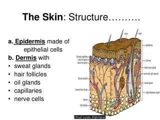
3. Dermis • a. Known as the inner, thicker portion of skin • b. Contains: • Blood vessels • Nerves • Nerve endings • Hair follicles • Sweat glands • Oil glands
c. Attached to an underlying layer of subcutaneous (fat) layer. • This fat layer helps absorb impacts, retain heat, store food.
Dermis • Second major skin region containing strong, flexible connective tissue • Cell types include fibroblasts, macrophages, and occasionally mast cells and white blood cells • Composed of two layers – papillary (fingerlike projections) and reticular
Layers of the Dermis: Papillary Layer • Papillary layer • Areolar connective tissue with collagen and elastic fibers • Its superior surface contains peglike projections called dermal papillae • Dermal papillae contain capillary loops, Meissner’s corpuscles, and free nerve endings
Layers of the Dermis: Reticular Layer • Reticular layer • Accounts for approximately 80% of the thickness of the skin • Collagen fibers in this layer add strength and resiliency to the skin • Elastin fibers provide stretch-recoil properties
Dermis Characteristics • Uneven surface • Contains: • Fingerlike papillae • Fibrous and connective tissue • Muscle fibers • Hair follicles • Blood vessels • Nerve fibers • Pacinian Corpuscles = heavy pressure • Meissner’s Corpuscles = light pressure -- Sevbaceous glands --Sweat glands
Sweat Glands • Different types prevent overheating of the body; secrete cerumen and milk • Eccrine sweat glands – found in palms, soles of the feet, and forehead • Apocrine sweat glands – found in axillary and anogenital areas • Ceruminous glands – modified apocrine glands in external ear canal that secrete cerumen • Mammary glands – specialized sweat glands that secrete milk
Sebaceous Glands • Simple alveolar glands found all over the body • Soften skin when stimulated by hormones • Secrete an oily secretion called sebum
Structure of a Nail • Scalelike modification of the epidermis on the distal, dorsal surface of fingers and toes
Hair • Filamentous strands of dead keratinized cells produced by hair follicles • Contains hard keratin which is tougher and more durable than soft keratin of the skin • Made up of the shaft projecting from the skin, and the root embedded in the skin • Consists of a core called the medulla, a cortex, and an outermost cuticle • Pigmented by melanocytes at the base of the hair
Hair Function and Distribution • Functions of hair include: • Helping to maintain warmth • Alerting the body to presence of insects on the skin • Guarding the scalp against physical trauma, heat loss, and sunlight • Hair is distributed over the entire skin surface except • Palms, soles, and lips • Nipples and portions of the external genitalia
Hair Follicle • Root sheath extending from the epidermal surface into the dermis • Deep end is expanded forming a hair bulb • A knot of sensory nerve endings (a root hair plexus) wraps around each hair bulb • Bending a hair stimulates these endings, hence our hairs act as sensitive touch receptors
Types of Hair • Vellus – pale, fine body hair found in children and the adult female • Terminal – coarse, long hair of eyebrows, scalp, axillary, and pubic regions
Hair Thinning and Baldness • Hair Thinning and Baldness • Alopecia – hair thinning in both sexes • True, or frank, baldness • Genetically determined and sex-influenced condition • Male pattern baldness – caused by follicular response to DHT
Functions of the Integumentary System • Protection – chemical, physical, and mechanical barrier • Body temperature regulation is accomplished by: • Dilation (cooling) and constriction (warming) of dermal vessels • Increasing sweat gland secretions to cool the body • Cutaneous sensation – exoreceptors sense touch and pain
Functions of the Integumentary System • Metabolic functions – synthesis of vitamin D in dermal blood vessels • Blood reservoir – skin blood vessels store up to 5% of the body’s blood volume • Excretion – limited amounts of nitrogenous wastes are eliminated from the body in sweat
Skin Cancer • Most skin tumors are benign and do not metastasize • A crucial risk factor for nonmelanoma skin cancers is the disabling of the p53 gene • Newly developed skin lotions can fix damaged DNA
Skin Cancer • The three major types of skin cancer are: • Basal cell carcinoma • Squamous cell carcinoma • Melanoma