Download
1 / 1
10 likes | 169 Vues
Methods* peripheral BS (16mL EDTA-BS) Ficoll-gradient centrifugation (650g, 20min) washing and centrifugation of the MNCs leukocyte labeling (40µl CD45 microbeads; 15min, 4°C) autoMACS (Miltenyi Biotec), deplete sensitive mode

E N D
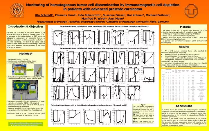
Methods* • peripheral BS (16mL EDTA-BS) • Ficoll-gradient centrifugation (650g, 20min) • washing and centrifugation of the MNCs • leukocyte labeling (40µl CD45 microbeads; 15min, 4°C) • autoMACS (Miltenyi Biotec), deplete sensitive mode • cytospin centrifugation of CD45- cell population (1 slide) collection of leukocytes (CD45+, DNA isolation) • immunocytochemistry (ICC: anti-CK-ab MNF116, Dako) • pathomorphological assessment:detection & quantification of disseminated tumor cells • comparison with clinical data of the affected patients • *References: Meye et al. Int J Oncol 21: 521-530 (2002) Schmidt et al. Int J Oncol in press MNC Up to 20 BS per day immuno-magnetic cell sorting (MACS) CD45+ CD45- labeled MNCsin 1 mL PBS Monitoring of hematogenous tumor cell dissemination by immunomagnetic cell depletion in patients with advanced prostate carcinoma Uta Schmidt1, Clemens Linné1, Udo Bilkenroth2 , Susanne Füssel1, Kai Krämer1, Michael Fröhner1, Manfred P. Wirth1, Axel Meye1 1Department of Urology, Technical University Dresden, 2Institute of Pathology, University Halle, Germany - 241 blood samples from 32 patients with advanced CaP (cT4) undergoing chemotherapy (median 6 per patient, range 3-15) - median age of the patients: 64.8 years (range 47.8-81.2) - median time of follow-up 67 weeks (range 16-120) - median serum PSA before chemotherapy 74.5ng/ml (range 1.4-2870.9 (determined by AxSym/Abbott ) - all patients were under hormonal ablation (LHRH analogons and antiandrogens) Introduction & Objectives Currently, the monitoring of therapeutic success in the palliative treatment of advanced prostate cancer (CaP) is restricted to the determination of PSA levels and radiographic assessment of metastatic lesions as objective parameters and the subjective evaluation of pain and well-being. The aim of our study was to assess whether the occurrence of circulating tumor cells might be an additional helpful parameter in the follow-up of cancer patients. Material Patients with tumor cells in their blood showing no PSA response during cytotoxic chemotherapy (Group 3) • Results • 77 of 241 samples contained tumor cells, classified by cytokeratin staining and/or morphology • a median of 5 cells per positive sample (range 1-862) • we could distinguish four groups of patients (figure) • no tumor cells were found in the samples of 5 out of 32 patients • for 13 patients, tumor cells were detectable in the peripheral blood before a PSA relapse occurred • one patient showed very high tumor cell numbers (more than 50 cells per slide, patient 1) at one time point of investigation • not all cells with an abnormal morphology expressed cytokeratin Patients with tumor cells in their blood showing a PSA response during cytotoxic chemotherapy (Group 4) Figure The X axis indicates the observation time in weeks, the left Y axis indicates the PSA levels, the right Y axis indicates the number of detected tumor cells Top: patients with tumor cells in their blood who did not show a temporary PSA decline of over 50% Middle: PSA responders with tumor cells Bottom: these patients had no detectable tumor cells in their blood Patients without tumor cells in their blood during cytotoxic chemotherapy (Groups 1 and 2) Conclusions In contrast to RT-PCR studies, the immunomagnetic enrichment strategy for the detection of disseminated tumor cells is suitable for a precise enumeration of the number of circulating tumor cells. Another advantage of this method is its independence of leukocyte counts for quantification. According to our results, tumor cell dissemination in late prostate cancer does not occur in a continuous fashion. In combination with the common follow-up parameters, tumor cell counts in the peripheral blood might be a useful measure to identify patients at higher risk for therapeutic failure. uta.schmidt2@mailbox.tu-dresden.de or www.tu-dresden.de\meduro\