Download
1 / 36
550 likes | 1.14k Vues
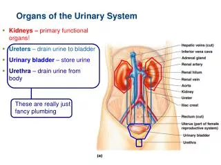
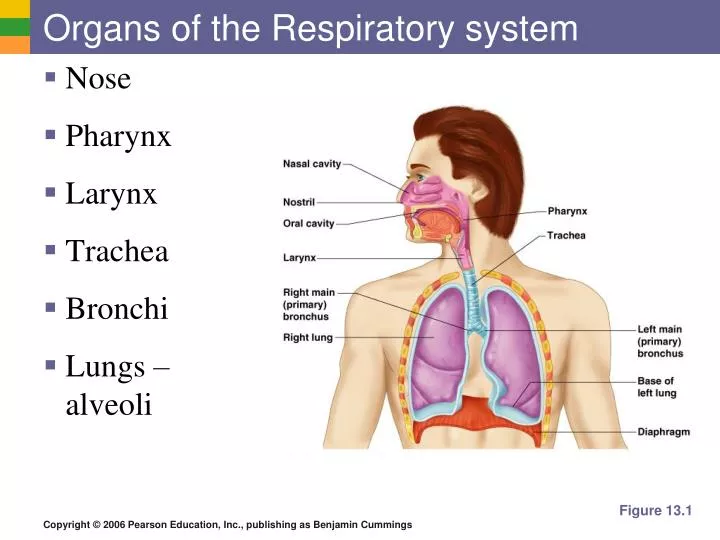
Organs of the Respiratory system. Nose Pharynx Larynx Trachea Bronchi Lungs – alveoli. Figure 13.1. Function of the Respiratory System. Oversees gas exchanges between the blood and external environment Exchange of gasses takes place within the lungs in the alveoli

E N D
Organs of the Respiratory system • Nose • Pharynx • Larynx • Trachea • Bronchi • Lungs – alveoli Figure 13.1
Function of the Respiratory System • Oversees gas exchanges between the blood and external environment • Exchange of gasses takes place within the lungs in the alveoli • Passageways to the lungs purify, warm, and humidify the incoming air
The Nose • The only externally visible part of the respiratory system • Air enters the nose through the external nares (nostrils) • The interior of the nose consists of a nasal cavity divided by a nasal septum
Upper Respiratory Tract Figure 13.2
Anatomy of the Nasal Cavity • Olfactory (smell) receptors are located in the mucosa on the superior surface • The rest of the cavity is lined with respiratory mucosa • Moistens air • Traps incoming foreign particles
Anatomy of the Nasal Cavity • The nasal cavity is separated from the oral cavity by the palate • Anterior hard palate (bone) • Posterior soft palate (muscle)
Paranasal Sinuses • Cavities within bones surrounding the nasal cavity • Frontal bone • Sphenoid bone • Ethmoid bone • Maxillary bone
Paranasal Sinuses • Function of the sinuses • Lighten the skull • Act as resonance chambers for speech • Produce mucus that drains into the nasal cavity
Pharynx (Throat) • Muscular passage from nasal cavity to larynx • The upper and middle pharynx are common passageways for air and food
Larynx (Voice Box) • Routes air and food into proper channels • Plays a role in speech • Made of eight rigid hyaline cartilages and a spoon-shaped flap of elastic cartilage (epiglottis)
Structures of the Larynx • Thyroid cartilage • Largest hyaline cartilage • Protrudes anteriorly (Adam’s apple) • Epiglottis • Superior opening of the larynx • Routes food to the larynx and air toward the trachea
Structures of the Larynx • Vocal cords (vocal folds) • Vibrate with expelled air to create sound (speech) • Glottis – opening between vocal cords
Trachea (Windpipe) • Connects larynx with bronchi • Lined with ciliated mucosa • Beat continuously in the opposite direction of incoming air • Expel mucus loaded with dust and other debris away from lungs • Walls are reinforced with C-shaped hyaline cartilage
Primary Bronchi • Formed by division of the trachea • Bronchi subdivide into smaller and smaller branches
Lungs • Occupy most of the thoracic cavity • Apex is near the clavicle (superior portion) • Base rests on the diaphragm (inferior portion)
Each lung is divided into lobes by fissures • Left lung – two lobes • Right lung – three lobes
Lungs Figure 13.4b
Coverings of the Lungs • Pulmonary (visceral) pleura covers the lung surface • Parietal pleura lines the walls of the thoracic cavity • Pleural fluid fills the area between layers of pleura to allow gliding
Bronchioles • Smallest branches of the bronchi • Terminal bronchioles end in alveoli Figure 13.5a
Capillaries Alveoli -Gas exchange takes place within the alveoli in the respiratory membrane -Pulmonary capillaries cover external surfaces of alveoli
Build a lung model • http://www.sciencefriday.com/program/archives/201006252 • Scientists building a real lung!
Gas Exchange • Gas crosses the respiratory membrane by diffusion • Oxygen enters the blood • Carbon dioxide enters the alveoli
Events of Respiration • External respiration – gas exchange between pulmonary blood and alveoli • Respiratory gas transport – transport of oxygen and carbon dioxide via the bloodstream • Internal respiration – gas exchange between blood and tissue cells in systemic capillaries
Gas Transport in the Blood • Oxygen transport in the blood • Inside red blood cells attached to hemoglobin (oxyhemoglobin [HbO2])
Mechanics of Breathing (Pulmonary Ventilation) • Two phases • Inspiration – flow of air into lung • Expiration – air leaving lung
Inspiration Diaphragm and intercostal muscles contract The size of the thoracic cavity increases External air is pulled not sucked into the lungs Figure 13.7a
Expiration • Largely a passive process which depends on natural lung elasticity • As muscles relax, air is pushed out of the lungs • Forced expiration can occur mostly by contracting internal intercostal muscles to depress the rib cage
Expiration Figure 13.7b
Nonrespiratory Air Movements • Can be caused by reflexes or voluntary actions • Examples • Cough and sneeze – clears lungs of debris • Laughing • Crying • Yawn • Hiccup
Respiratory Sounds • Sounds are monitored with a stethoscope • Bronchial sounds – produced by air rushing through trachea and bronchi • Vesicular breathing sounds – soft sounds of air filling alveoli
Factors Influencing Respiratory Rate and Depth • Physical factors • Increased body temperature • Exercise • Talking • Coughing • Volition (conscious control) • Emotional factors
Emphysema • Alveoli enlarge as adjacent chambers break through • Chronic inflammation promotes lung fibrosis • Airways collapse during expiration • Patients use a large amount of energy to exhale • Overinflation of the lungs leads to a permanently expanded barrel chest
Chronic Bronchitis • Mucosa of the lower respiratory passages becomes severely inflamed • Mucus production increases • Pooled mucus impairs ventilation and gas exchange • Risk of lung infection increases • Pneumonia is common
Lung Cancer • Accounts for 1/3 of all cancer deaths in the United States • Increased incidence associated with smoking • Three common types • Squamous cell carcinoma • Adenocarcinoma • Small cell carcinoma
Sudden Infant Death syndrome (SIDS) • Apparently healthy infant stops breathing and dies during sleep • Some cases are thought to be a problem of the neural respiratory control center • One third of cases appear to be due to heart rhythm abnormalities
Asthma • Chronic inflamed hypersensitive bronchiole passages • Response to irritants with coughing, and wheezing