Download
1 / 50
500 likes | 608 Vues
Sicklecell Anemia - A Disease of Diverse Populations. Jennie Aizenman Scott Bronson Uwe Hilgert. Sickle Cell Anemia. Red blood cells shaped like sickles. A genetic disorder: HBB A , HBB S Severe if both copies of gene are affected. Benign if only one copy of gene is affected.

E N D
Sicklecell Anemia - A Disease of Diverse Populations Jennie Aizenman Scott Bronson Uwe Hilgert
Sickle Cell Anemia Red blood cells shaped like sickles. A genetic disorder: HBBA, HBBS Severe if both copies of gene are affected. Benign if only one copy of gene is affected.
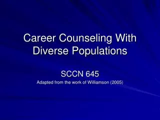
Why does disease persist? HH – healthy HS – benign SS – severe
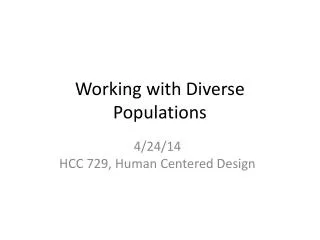
Malaria prevalence HBS Allele Mosquito (Anopheles species)
Genes and Life How DNA Encodes Life DNA RNA Protein DNA - What are genes? Transcription – Copying the code Translation – Reading the code Code Table - The Code
What Are Proteins? Polymers of amino acids 20 different amino acids Consist of core (-NH-CO-) and side group. 20 side groups 20 characteristics • Go to http://info.bio.cmu.edu/Courses/Bioche mMols/AAViewer/AAVFrameset.htm • Set left molecule to alanine. • Set right molecule to tryptophane. • How do the two amino acids differ? • Examine glutamate, proline, valine.
How Do Proteins Work? Proteins: Structure, Catalysts, Sensors, … Proteins are polymers (long chains) of amino acids. Sequence Structure Function Let’s look at a protein together.
Bioinformatics DNA and protein sequences are stored in databases. Bioinformatics provides the tools to handle and analyze these data. Bioinformatics is the science of unraveling the information in biological molecules utilizing computer technology.
Bioinformatics: Structures in Databases NCBI: National Center for Biotechnology Information Open browser, go to http://www.ncbi.nlm.nih.gov/ • Find Search Entrez for. • Change Entrez to Structure. • Type hemoglobin (after for). • Click Go. • Find entry 1KOY, click it, click View 3D Structure.
Hemoglobin • Oxygen is picked up by iron (Fe) • which is held by porphyrine (ring), • which is presented by protein chain, • of which 4 aggregate in Hb molecule, • so that together (cooperatively) they influence irons affinity to oxygen
Allosteric Effect - Cooperativity http://www.rcsb.org/pdb/molecules/pdb41_2.html Hemoglobin’s 576 amino acids determine its ability to bind and release oxygen and serve as oxygen transport vehicle in red blood cells.
Find the Genes GenBank database maintained by the National Center for Biotechnology Information (NCBI) Go to http://www.ncbi.nlm.nih.gov/ Which organisms can you find mentioned at the NCBI homepage? What other things catch your attention?
“Roadmaps” for Genomes Map Viewer: tool to locate genes • Go to http://www.ncbi.nlm.nih.gov/. • Find Map Viewer, click on it. • Which genomes can you access? • Click homo sapiens. • Answer the questions on your worksheet.
Search the Map Find hemoglobin gene(s) • Find Search for. • Into the search window type hemoglobin. • Click Find. • Answer the questions in the worksheet.
Find the Hemoglobin Beta Gene (HBB) • Click on HBB. • Answer the questions on the worksheet.
Find the HBB Nucleotide Sequence • Go to http://www.ncbi.nlm.nih.gov/. • Find Search Entrez for. • Delete Entrez. • Behind for write hemoglobin homo sapiens. • Click Go. • Answer the questions in the worksheet.
Find and Examine the Nucleotide Sequence • Click Nucleotide: sequence database (GenBank). • Click Homo sapiens hemoglobin, beta (HBB) mRNA. • Click on NM_000518. • Answer the questions on the worksheet.
Work With the Nucleotide Sequence Transfer RNA sequence to database • Highlight and copy nucleotide 1 through 626. • Go to http://www.bioservers.org/bioserver/ • Under SequenceServer click Enter. • Click CREATE SEQUENCE. • Paste the sequence into the big window. • Write HBB mRNA into the Name window. • Click OK. • This is the RNA sequence, it’s 626 bp long.
Find the Gene in the Human DNA Sequence • How? • What’s the difference between mRNA and a gene? • So, how does having mRNA help you find its gene in DNA?
Align mRNA with DNA to Identify Gene Structure • Copy HBBcDNA from SequenceServer. • Open a second browser window. • Go to SIM 4: http://pbil.univ-lyon1.fr/sim4.php. • Paste the sequence into the cDNA window. • In SequenceServer, change HBB cDNA to HBB gene. • Transfer sequence into the lower window of the SIM4 tool. • Click Submit. • Save result onto desktop and visualize the alignment with LalnView.
How Can Genes Cause Disease? What is a genetic disorder? • An inheritable disorder. • Discuss healthy vs. carrier vs. disease. • Beta globin gene: HbbA (normal gene/protein) • Sickle cell gene: HbbS (mutated/faulty gene/protein) • Healthy people: HbbA HbbA • Sickle cell trait: HbbA HbbS (carrier, sickle cell disease) • Sickle cell anemia: HbbS HbbS
Find the Mutation in the Gene • How? What’s a mutation? • Align the sequences in SequenceServer. • Go to http://www.bioservers.org/. • Change Classes to Public. • Find HBB and Sickle Cell Anemia. • On the left, check the box. • On the bottom, click OK.
Find HBB cDNA, homo sapiens. • What does cDNA stand for? • Find the word None, click it. • Click HBS CDS, homo sapiens. • What does CDS stand for? • Find COMPARE. • Set box next to it to Align Clustal W. • Click COMPARE – WAIT!!
Find Mutations • Mutations are changes in sequences. • Find the differences between the HBB and HBS nucleotide sequences. • Answer the questions in the worksheet.
Find Mutations • How? • Identify what changes the nucleotide differences cause within the protein. • First translate the HBS and HBB coding DNA into their respective amino acid sequences. • How? • Code Table - The Code
How do the DNA mutations affect the protein? Translate DNA into amino acids. • Click Open for HBB cDNA, homo sapiens. • Move the cursor just before the A of the ATG on the third line. • Hit Return/Enter on your keyboard, this moves the ATG to the fourth line. • Highlight and copy the sequence from the ATG to the end (don’t worry about the stop codon …). • Click Done.
How do the DNA mutations affect the protein? Translate DNA into aminoacids. • Open a new browser. • Open Gene Boy http://www.dnai.org/geneboy/. • Click Your Sequence. • Paste the sequence into the workspace. • Click Save Sequence (should yield 576 nucleotides).
How do the DNA mutations affect the protein? Translate DNA into amino acids. • On the Operations panel to the right click Transform Sequence. • Select Amino Acids.. • Highlight the sequence under Reading Frame RF1 and copy it. • Open the Word program and paste the amino acid sequence into it. • Place a carriage return at the end of the sequence. • Place a “>” sign in front of the sequence, followed by the letters “HBB”. • Type a carriage return.
How do the DNA mutations affect the protein? • Repeat the process for the sickle cell mRNA (HBS CDS, homo sapiens) with the following modifications: • Use sequence from HBS CDS, homo sapiens instead of HBB cDNA, homo sapiens; • copy the entire sequence; • pasting this sequence into GeneBoy should yield you 444 amino acids; • write HBS before the sequence instead of HBB.
How do the DNA mutations affect the protein? Align the amino acid sequences for HBB and HBS. • Highlight and copy the content of the Word-file. • Go to http://www.ebi.ac.uk/clustalw/. • Find Enter or Paste a set of Sequences … and paste the sequence into the box. • Click Run. • WAIT!
How do the DNA mutations affect the protein? • The result window shows an alignment of the two amino acid sequences. • Underneath the alignment is a string of stars denoting identical amino acids. Find the amino acid differences between HBB and HBS. Ignore, however, the end where only HBB shows amino acids; this region is not part of the HBB protein. The HBB as well as the HBS proteins end with the amino acid sequence AHKYH. • What are the differences between HBB and HBS?
Effect of the Glu Val mutation on HBB Structure How do valine and glutamate differ? • http://info.bio.cmu.edu/Courses/BiochemMols/AAViewer/AAVFrameset.htm. • Set left window to valine. • Turn the molecule so that the aminoacid-core molecules, the red/blue “V”, is positioned on top. • Set the right window to glutamate; position red/blue “V” on top. • Compare and contrast the two structures. • Answer the questions in the worksheet.
Effect of the Glu Val mutation on HBB Structure How do HBB and HBS differ? • Open http://www.dnalc.org/bioinformatics/sickle_cell_module/hbb_cn3d.val. • Open http://www.dnalc.org/bioinformatics/sickle_cell_module/hbbs_cn3d.val. • Close the Message Log windows. • Align the Cn3D windows side by side. • Place each Sequence windows underneath its structure.
Effect of the Glu Val mutation on HBB Structure How do HBB and HBS differ? • Can you see any difference between HBB and HBS? • Highlight the differences: E in HBB and V in HBS. • Orient highlighted amino acids in similar positions. • Can you identify differences now? • Go to Style, Rendering Shortcuts, Worms. • Zoom in on the highlighted amino acids. • Go to Style, Rendering Shortcuts, Toggle Sidechains. • Go to Style, Coloring Shortcuts, Structure. • What difference do you see between HBB and HBS?
Align the HBS and HBB Proteins Go to NCBI at http://www.ncbi.nlm.nih.gov/. • Find the words Search Entrez for. • Change Entrez to Structure. • Into the search window type HBS; hit Go. • Click on 2HBS. • Click on the term Chain B (find the blue bar …). • Click on View 3D Structure. • Click on Open. • Maximize the Cn3D screen; align the Sequence screen underneath; close the Message Log screen.
Align the HBS and HBB Proteins • How different are the two proteins? • Identify and highlight in both sequences the amino acid that’s different. • Can you see a difference now? • Go to Style, Rendering Shortcuts, click Toggle Sidechains. Make sure the V and E in position 6 of both sequences are highlighted. • Can you see a difference now? • Change the highlighting from position 6 to position 5 (Proline; P). • Can you see a difference now?
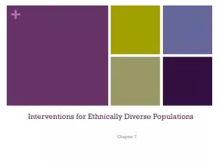
Summary Glutamic Acid, E Hydrophilic, Polar Valine, V Hydrophobic, Non-polar
http://www.ygyh.org/sickle/have02.htm http://www.ygyh.org/sickle/have03.htm http://www.ygyh.org/sickle/have07.htm
Malaria Caused by protozoa of the Plasmodium genus. Overlap of malaria endemic areas (Plasmodium falciparum) and areas where sickle cell mutation occurs. Sickle cell mutation confers protection from Plasmodium falciparum. Anopheles mosquito
Malaria infects 300 to 500 million people worldwide and accounts for over 1 million deaths annually.