Download
1 / 1
10 likes | 89 Vues
3D Imaging of Dendritic Cells Using Serial Ion-Abrasion Scanning Electron Microscopy (SIA-SEM) Reveals That HIV-1 is Stored Within Surface-connected Membrane Folds. N. Avishai*, K. Olszens***, A. Avishai*, M. Hitomi**, H. J. Yu***, A.H. Heuer* and D. McDonald***

E N D
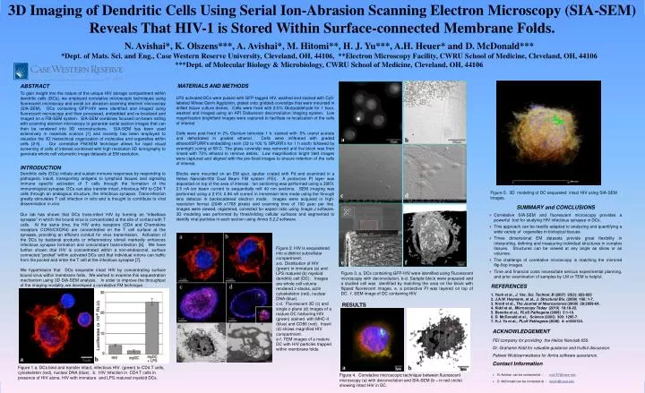
3D Imaging of Dendritic Cells Using Serial Ion-Abrasion Scanning Electron Microscopy (SIA-SEM) Reveals That HIV-1 is Stored Within Surface-connected Membrane Folds. N. Avishai*, K. Olszens***, A. Avishai*, M. Hitomi**, H. J. Yu***, A.H. Heuer* and D. McDonald*** *Dept. of Mats. Sci. and Eng., Case Western Reserve University, Cleveland, OH, 44106, **Electron Microscopy Facility, CWRU School of Medicine, Cleveland, OH, 44106 ***Dept. of Molecular Biology & Microbiology, CWRU School of Medicine, Cleveland, OH, 44106 ABSTRACT To gain insight into the nature of the unique HIV storage compartment within dendritic cells (DCs), we employed correlative microscopic techniques using fluorescent microscopy and serial ion abrasion scanning electron microscopy (SIA-SEM). DCs containing GFP-HIV were identified and imaged using fluorescent microscopy and then processed, embedded and re-localized and imaged on a FIB-SEM system. SIA-SEM combines focused ion-beam milling with scanning electron microscopy to generate serial section images that can then be rendered into 3D reconstructions. SIA-SEM has been used extensively in materials science [1] and recently has been employed to visualize the 3D hierarchical organization of molecules and organelles within cells [2-5]. Our correlative FM/SEM technique allows for rapid visual screening of cells of interest combined with high resolution 3D tomography to generate whole cell volumetric image datasets at EM resolution. INTRODUCTION Dendritic cells (DCs) initiate and sustain immune responses by responding to pathogenic insult, transporting antigens to lymphoid tissues and signaling immune specific activation of T cells through the formation of the immunological synapse. DCs can also transfer intact, infectious HIV to CD4 T cells through an analogous structure, the infectious synapse. Trans-infection greatly stimulates T cell infection in vitro and is thought to contribute to viral dissemination in vivo. Our lab has shown that DCs trans-infect HIV by forming an “infectious synapse” in which the bound virus is concentrated at the site of contact with T cells. At the same time, the HIV entry receptors (CD4 and Chemokine receptors CCR5/CXCR4) are concentrated on the T cell surface at the synapse, providing an efficient conduit for virus transmission. Activation of the DCs by bacterial products or inflammatory stimuli markedly enhances infectious synapse formation and concomitant trans-infection [6]. We have further shown that HIV is concentrated within a non-endosomal, surface connected “pocket” within activated DCs and that individual virions can traffic from the pocket and enter the T cell at the infectious synapse [7]. We hypothesize that DCs sequester intact HIV by concentrating surface bound virus within membrane folds. We wished to examine this sequestration mechanism using 3D SIA-SEM analysis. In order to improve the throughput of the imaging modality, we developed a correlative FM technique. MATERIALS AND METHODS LPS activated DCs were pulsed with GFP-tagged HIV, washed and stained with Cy5-labeled Wheat Germ Agglutinin, plated onto gridded coverslips that were mounted in drilled tissue culture dishes. Cells were fixed with 2.5% Glutaraldehyde for 1 hour, washed and imaged using an API Deltavisiondeconvolution imaging system. Low magnification brightfieldimages were captured to facilitate re-localization of the cells of interest. Cells were post-fixed in 2% Osmium tetroxide 1 h, stained with .5% uranyl acetate and dehydrated in graded ethanol. Cells were infiltrated with graded ethanol/SPURR’s embedding resin (33 to 100 % SPURR’s for 1 h each) followed by overnight curing at 68˚C. The glass coverslip was removed and the block was then rinsed with 70% ethanol to remove debris. Low magnification bright field images were captured and aligned with the pre-fixed images to ensure retention of the cells of interest. Blocks were mounted on an EM spur, sputter coated with Pd and examined in a Helios Nanolab-650 Dual Beam FIB system (FEI). A protective Pt layer was deposited on top of the area of interest. Ion sectioning was performed using a 30KV, 2.5 nA ion beam current to sequentially mill 40 nm sections. SEM imaging was performed using a 2 KV, 0.84 nA current in immersion lens mode using the through lens detector in backscattered electron mode. Images were acquired in high-resolution format (2048 x1768 pixels) and scanning time of 100 µsec per line. Images were viewed, registered, corrected for aspect ratio using Image J software. 3D modeling was performed by thresholding cellular surfaces and segmented to identify viral particles in each section using Amira 5.2.2 software. a b Figure 5. 3D modeling of DC sequested intact HIV using SIA-SEM images. c d • SUMMARY and CONCLUSIONS • Correlative SIA-SEM and fluorescent microscopy provides a powerful tool for studying HIV infectious synapse in DCs. • This approach can be readily adapted to analyzing and quantifying a wide variety of organelles in biological tissues. • Three dimensional EM datasets provide great flexibility in interpreting, defining and measuring individual structures in complex tissues. Structures can be viewed at any angle as slices or as volumes. • The challenge of correlative microscopy is matching the mirrored flip-flop images. • Time and financial costs necessitate serious experimental planning, and prior examination of samples by LM or TEM is helpful. Figure 2. HIV is sequestered into a distinct subcellular compartment. a-b. Distribution of HIV (green) in immature (a) and LPS matured (b) myeloid dendritic cell (DC). Images are whole cell volume rendered z-stacks, actin cytoskeleton (red), nuclear DNA (blue). c-d. Fluorescent 3D (c) and single z-plane (d) images of a mature DC harboring HIV (green) stained with MHC-II (blue) and CD86 (red). Insert (d) shows magnified HIV compartment. e-f. TEM images of a mature DC with HIV particles trapped within membrane folds. e f Figure 3. a. DCs containing GFP-HIV were identified using Fluorescent microscopy with deconvolution. b-d. Sample block were prepared and a studied cell was identified by matching the area on the block with flipped fluorescent images. e. a protective Pt was layered on top of DC. f. SEM image of DC containing HIV. REFERENCES 1. Yeoh et al., J. Vac. Sci. Technol. B (2007) 25(3): 922-925 2. J.A.W. Heymann, et al., J. Structural Bio. (2009) 166: 1-7. 3. Knott et al., The Journal of Neuroscience (2008) 28:2959-64. 4. Kidd et al., Microscopy Today (2010) 18:18-22. 5. Benette et al., PLoS Pathogens (2009) 5:1-10. 6. D. McDonald et al., Science (2003) 300: 1295-7. 7. H.J. Yu et al., PLoS Pathogens (2008) 4: e1000134. RESULTS • ACKNOWLEDGEMENT • FEI company for providing the Helios Nanolab 650. • Dr. Grahame Kidd for valuable guidance and fruitful discussion. • PatiwetWuttisarnwattana for Amira software assistance. • Contact Information • N. Avishai can be contacted at : nxa157@case.edu • D. McDonald can be contacted at : djm41@case.edu a b a b Figure 1 a. DCs bind and transfer intact, infectious HIV (green) to CD4 T cells, cytoskeleton (red), nuclear DNA (blue). b. HIV Infection in CD4 T cells in presence of HIV alone, HIV with immature and LPS matured myeloid DCs. Figure 4. Correlative microscopic technique between fluorescent microscopy (a) with deconvolution and SIA-SEM (b – in red circle) showing intact HIV in DC.