Download
1 / 64
860 likes | 1.84k Vues
Regional anatomy of neck. 山东大学医学院 解剖教研室 李振华. Parts and regions of the neck. Boundaries Superior - a line joining inferior border of mandible, angle of mandible, tip of mastoid process, superior nuchal line and external occipital protuberance

E N D
Regional anatomy of neck 山东大学医学院 解剖教研室 李振华
Parts and regions of the neck Boundaries • Superior- a line joining inferior border of mandible, angle of mandible, tip of mastoid process, superior nuchal line and external occipital protuberance • Inferior- a line joining jugular notch, sternoclavicular joint, superior border of clavicle, acromion and spinous processes of C7
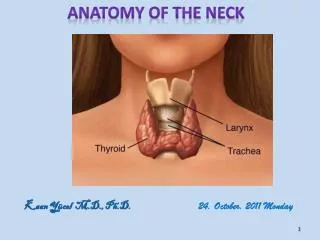
Landmarks of the neck • Hyoid bone 舌骨 • Thyroid cartilage 甲状软骨 • Cricoid cartilage 环状软骨 • Catotid tubercle 颈动脉结节
Landmarks of the neck • Hyoid bone 舌骨 • Thyroid cartilage 甲状软骨 • Cricoid cartilage 环状软骨 • Catotid tubercle 颈动脉结节
Landmarks of the neck • Sternocleidomastoid • Suprasternal fossa • Greater supraclaviclar fossa
Regions of neck • Neck颈 • Anterior region of neck 颈前区 • Sternocleidomastoid region 胸锁乳突肌区 • Lateral region of neck 颈外侧区 • Nape项部
Triangles of anterior region of neck • Suprahyoid region舌骨上区 • Submental triangle 颏下三角 • Submandibular triangle下颌下三角 • Infrahyoid region舌骨下区 • Carotid triangle 颈动脉三角 • Muscular triangle 肌三角
Triangles of lateral region of neck • Occipital triangle 枕三角 • supraclavicular triangle 锁骨上三角 (greater supraclavicular fossa)锁骨上大窝
Skin of the neck • The natural line of cleavage of the skin are constant and run almost horizontally around the neck
Superficial fascia Contents • Platysma 颈阔肌 • Superficial veins • Anterior jugular v. 颈前静脉 • External jugular v. 颈外静脉 • Cutaneous nerves • Lesser occipital n. 枕小神经 • Greet auricular n. 耳大神经 • Transverse nerve of neck 颈横神经 • Supraclavicular n. 锁骨上神经 • Cervical branch of facial n. 面神经颈支
★Cervical fascia颈筋膜 Superficial layerof cervical fascia颈筋膜浅层(investing fascia 封套筋膜) • Encloses trapezius, sternocleidomastoid, posterior belly of digastric and parotid and submandibular glands • Attached to bony landmarks of upper and lower boundaries of neck and zygomatic arch of face
★ Cervical fascia颈筋膜 Pretracheal layer气管前层 • Lies deep to the infrahyoid muscle • Encloses viscera of neck: pharynx, larynx, trachea, esophagus, thyroid gland and parathyroid glands • Completely surrounds thyroid gland, forming a sheath for it, and bind the gland to larynx to form suspensory ligament of thyroid gland甲状腺悬韧带 • Extends from arch of cricoid cartilage, thyroid cartilage and hyoid bone to fibrous pericardium of superior mediastinum
★ Cervical fascia颈筋膜 Prevertebral layer椎前层 • Lies anterior to bodies of cervical vertebrae and prevertebral muscles; extends from base of skull downward into the superior mediastinum, continuous with anterior longitudinal lig. and endothoracic fascia • Covers subclavian vessels and roots of brachial plexus • Extends into upper limb as axillary sheath
★ Cervical fascia颈筋膜 Carotid sheath颈动脉鞘 • Formed by components of all three layers of deep cervical fascia • Contains common and internal carotid arteries, internal jugular vein, and vagus nerve
Fascia spaces筋膜间隙 Suprasternal space胸骨上间隙 • 3-4cm above manubrium of sterni the investing fascia splits into two layers, which are attached to the anterior and posterior margins of the upper border of the manubrium, between these two layers is a slit-like space, called the suprasternal space • Contains connective tissue, and sometimes a lymph node
fascia spaces筋膜间隙 Pretracheal space气管前间隙 • Lies between pretracheal layer and cervical part of trachea • Contains arteria thyroidea ima, inferior thyroid v., unpaired thyroid venous plexus, brachiocephalic trunk and left brachiocephalic v.
Fascia spaces筋膜间隙 Retropharyngeal space咽后间隙 • Lies between prevertebral layer and buccopharyngeal fascia Prevertebral space椎前间隙 • Lies between prevertebral muscles, cervical part of vertebral column and prevertebral layer
Suprahyoid region Submental triangle 颏下三角 • Lies below the chin • Boundaries • Laterally by anterior bellies of digastric • Inferiorly by the body of hyoid bone • Covered by skin, superficial fascia and investing fascia • Floor-mylohyoid muscles • Contents-submental lymph nodes
Suprahyoid region Submandibular triangle下颌下三角 • Boundaries • Anterior and posterior bellies of digastric • Lower border of the body of the mandible • Covered by skin, superficial fascia, platysma and investing fascia • Floor- mylohyoid, hyoglossus and middle constrictor of pharynx • Contents-submandibular gland, facial a., v., hypoglossal n. lingual a. v. and n., submandibular ganglion and submandibular lymph nodes
Infrahyoid region ★ Carotid triangle颈动脉三角 • Boundaries • Anterior border of sternocleidomastoid • Superior belly of omohyoid • Posterior belly of digastic • Covered by skin, superficial fascia, platysma and investing fascia • Deep-prevertebral fascia • Medial - lateral wall of pharynx
Infrahyoid region ★ Carotid triangle颈动脉三角 • Contents • Common carotid a. and its branches • Internal jugular v. and its tributaries • Hypoglossal n. with its descending branches • Vagus nerve • Accessory nerve • Deep cervical lymph nodes
Infrahyoid region Ralations of posterior belly of digastic • Superficial • great auricular n. • retromandibular v. • cervical branch of facial n. • Deep • internal and external carotid a. • internal jugular v. • Ⅹ~Ⅻ cranial n. • cervical part of sympathetic trunk • Superiorly • posterior auricular a. • facial a. • glossopharyngeal n. • Infeiorly • occipital a. • hypoglossal n.
Infrahyoid region ★Muscular triangle肌三角 • Bounded by midline of the neck, superior belly of the omohyoid and anterior border of the sternocleidomastoid. • Covered by skin, superficial fascia, platysma, anterior jugular v., coutaneous n. and investing fascia • Deep-prevertebral fascia
Infrahyoid region Muscular triangle肌三角 • Contents • Superior belly of omohyoid • Sternohyoid • Sternothyroid • Thyrohyoid • Thyroid gland • Parathyroid gland • Cervical part of trachea and esophagus
★Thyroid gland 甲状腺 Shape and position • H-shape • Left and right lobes: lie on either side of inferior part of larynx and superior part of trachea, extend from middle of thyroid cartilage to level of sixth trachea cartilage • Isthmus: overlies 2nd to 4th tracheal cartilage • Pyramidal lobe: some times arises from isthmus
★ Thyroid gland 甲状腺 Coverings of the thyroid gland • False capsule: a sheath of pretracheal fascia which is attached to arch of cricoid and thyroid cartilages to form the suspensory ligament of thyroid gland, hence, the thyroid gland moves with larynx during swallowing and oscillates during speaking • True capsule: fibrous capsule • Space between sheath and capsule of thyroid gland: there are loose connective tissue, vessels, nerves and parathyroid glands
★ Thyroid gland 甲状腺 Relations of the thyroid gland • Anteriorly: • Skin • superficial fascia • investing fascia • Infrahyoid muscles and pretracheal fascia • Posteromedially: • Larynx and trachea • Pharynx and esophagus • Recurrent laryngeal nerve • Posterolaterally: • Carotid sheath with common carotid a., internal jugular v., and vagus n. • Cervical sympathetic trunk
★Arteries of the thyroid gland Superior thyroid a. 甲状腺上动脉 • Branch of external carotid a. • Runs superficial and parallel to the external branch of superior laryngeal n. to reach the upper pole of thyroid gland • Gives off superior laryngeal a. in company with internal branch of superior laryngeal n.
★Arteries of the thyroid gland Inferior thyroid artery甲状腺下动脉 • Branch of thyrocervical trunkof subclavian a. • Turns medially and downward, reaches the posterior border of the thyroid gland, where it is closely related to the recurrent laryngeal n. • Supplies inferior pole of thyroid gland
★ Arteries of the thyroid gland Arteria thyroidea ima 甲状腺最下动脉 May arise (4%) from the brachiocephalic a. or aortic arch
★Nerves of the larynx Superior laryngeal n.喉上神经 • Internal branch 内支:which pierces thyrohyoid membrane to innervates mucous membrane of larynx above fissure of glottis • External branch外支:is fine n., which descends in company with the superior thyroid a. and supplies cricothyroid
★Nerves of the larynx Recurrent laryngeal nerves喉返神经 • Ascend in tracheo-esophageal groove • Pass deep to the lobe of the thyroid gland and come into close relationship with the inferior thyroid a. • Cross either in front of or behind the artery of may pass between its branches • Nerves enter larynx posterior to cricothyroid joint, the nerve is now called inferior laryngeal nerve • Innervations: laryngeal mucosa below fissure of glottis , all laryngeal laryngeal muscles except cricothyroid
Venous drainage of the thyroid gland • Superior thyroid veins drain into internal jugular vein • Middle thyroid veins drain into internal jugular vein • Inferior thyroid veins of two sides anastomose with one another as they descend in front of the trachea to form unpaired thyroid venous plexus 甲状腺奇静脉丛. They drain into brachiocephalic veins.
★ Parathyroid gland 甲状旁腺 • Yellowish-brown, ovoid bodies • Position • Two superior parathyroid glands: lie at junction of superior and middle third of posterior border of thyroid gland • Two inferior parathyroid glands: lie near the inferior thyroid artery, close to the inferior poles of thyroid gland • Function: regulate calcium and phosphate balance and is therefore essential for life
Cervical part of trachea 气管颈部 • Begins at lower end of larynx-level of C6 vertebra • Consists of a series of incomplete cartilage rings • Extends into thorax
Relations of cervical part of trachea 气管颈部 ★ Anteriorly • Skin • Superficial fascia • Investing fascia • Suprasternal space and jugular arch • Infrahyoid muscles and pretracheal fascia • Isthmus of thyroid gland ( in front of the 2nd to 4th tracheal cartilage) • Inferior thyroid v. and unpaired thyroid venous plexus • Arteria thyroid ima ( if present) • Thymus, left brachiocephalic v. and aortic arch in child
Relations of cervical part of trachea 气管颈部 • Superolaterally • lobes of the thyroid gland ( down as far as the sixth ring) • Posteriorly • Esophagus • R. & L. recurrent laryngeal nerves • Posterlaterally • Cervical sympathetic trunk • Carotid sheath
Cervical part of esophagus 食管颈部 • Extending from pharynx at level of C6 vertebra • Descends through the neck, it inclines to the left side • Relations of the cervical part of esophagus • Anteriorly • Trachea • Recurrent laryngeal nerves • Posteriorly • Prevertebral layer of cervicl fascia • Longus colli • Vertebral column • Laterally • Lobe of the thyroid gland • Carotid sheath with common carotid a., internal jugular v., and vagus n.
Sternocleidomastoid region 胸锁乳突肌区 • Covered by sternocleidomastoid • Contents • Ansa cervicalis • Carotid sheath • Cervical plexus • Cervical part of sympathetic trunk
Root of neck颈根部 • At thoracic inlet • Formed by • Anteriorly-manubrium sterni • Posteriorly-body of first thoracic vertebra • Laterally-first rib and costal cartilage • Central markers-scalenus anterior
Root of neck颈根部 Contents • Cupula of pleura-extends up into the neck, over the apex of lung, 2~3cm above the medial third of clavicle • Subclavian v. • Thoracic duct and right lymphatic duct • Subclavian a. • Vagus n. • Phrenic n.
Triangle of the vertebral a. 椎动脉三角 • Boundaries • Medially-longus colli • Laterally-scalenus anterior • Inferiorly-first part of subclavian a. • Apex-transverse process of C6 • Posteriorly-cupula of pleura, transverse process of C7, anterior rami of C8 spinal nerves, costal neck of 1st rib • Anteriorly-carotid sheath, phrenic n. and arch of thoracic duct (left) • Contents • Vertebral a. and v. • Inferior thyroid a. • Cervical part of sympathetic trunk • Cevicothoracic ganglion
Lateral region of neck 颈外侧区 • Bounded by posterior border of sternocleidomastoid, anterior border of trapezius and middle third of clavicle • Divided by inferior belly of omohyoid into occipital triangle and supraclavicular triangle
Occipital triangle 枕三角 • Bounded by posterior border of sternocleidomastoid, anterior border of trapezius and superior border of inferior belly of omohyoid • Covered by skin, superficial fascia, and investing fascia • Deep-prevertebral fascia and scalenus anterior, scalenus medius, scalenus posterior, splenius capitis and levator scapulae • Conents • Accessory n.-emerges above the middle of the posterior border of sternocleidomastoid and crosses the occipital triangle to trapezius • Cervical and brachial plexuses
Supraclavicular triangle 锁骨上三角 • Bounded by posterior border of sternocleidomastoid, inferior belly of omohyoid and middle third of clavicle • Covered by skin, superficial fascia, and investing fascia • Deep-prevertebral fascia and inferior parts of scalenus • Conents • Subclavian v. and venous angle • Subclavian a. • Brachial plexus
Skin incisions • Make the skin incisions shown in figure • Reflect the skin posteriorly to well behind the ear.
Dissection of Superficial Structures • Note the underlying platysma muscle, a muscle of facial expression, which has migrated onto the neck. Beneath the platysma lie the supraclavicular cutaneous nerves (C3-4) (medial,intermediate and lateral). Slightly superior to the middle of the posterior border of the sternocleidomastoid muscle, locate the spinal accessory nerve coursing downward toward the trapezius muscle. Platysma
Dissection of Superficial Structures • Using your scissors incise and spread the tough fascial covering of the posterior triangle and locate the lesser occipital nerve (C2-3) emerging close to CN.Ⅺ, note the direction that each nerve takes as it traverses the posterior triangle. • Next locate the great auricular nerve (C2-3) which ascends posterior and parallel with the external jugular vein on the sternoclidomastoid. • Try to identify the small transverse cervical nerve (C2-3) supplying skin over the anterior neck. • Look for the facial vein, retromandibular vein and, if present, the small anterior jugular vein, and review the external jugular system.
Lesser occipital n. External jugular vein Greet auricular n. Transverse nerve of neck Supraclavicular n. Anterior jugular vein Cutaneous nerves and superficial veins