Download
1 / 37
790 likes | 3.43k Vues
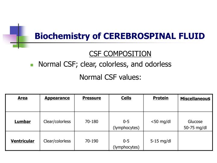
Biochemistry of CEREBROSPINAL FLUID. CSF COMPOSITION Normal CSF; clear, colorless, and odorless. Normal CSF values:. CEREBROSPINAL FLUID. CLINICAL CONSIDERATIONS Noncommunicating (obstructive) hydrocephalus occurs more frequently CSF of ventricles unable to reach subarachnoid space

E N D
Biochemistry of CEREBROSPINAL FLUID CSF COMPOSITION • Normal CSF; clear, colorless, and odorless Normal CSF values:
CEREBROSPINAL FLUID CLINICAL CONSIDERATIONS • Noncommunicating (obstructive) hydrocephalus occurs more frequently • CSF of ventricles unable to reach subarachnoid space • Production of CSF continues • Gyri are flattened against inside of skull • If skull is still pliable head may enlarge
CEREBROSPINAL FLUID CLINICAL CONSIDERATIONS • Communicating hydrocephalus; obstruction is in subarchnoid space due to thickening of the arachnoid with resultant block of return-flow channels • Can be the result of prior bleeding or meningitis • If ICP is increased due to excess CSF, central canal of spinal cord may dilate
CEREBROSPINAL FLUID CLINICAL CONSIDERATIONS • Various procedures have been developed to bypass the obstruction in noncommunicating hydrocephalus or to improve overall absorption in general
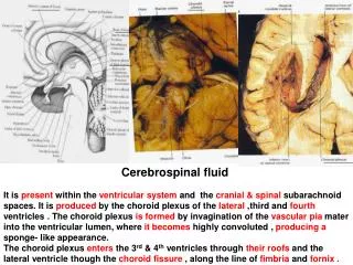
CIRCULATION OF CSF Circulation: CSF is mainlyformed in choroid pleaxus of the lateral ventricle. CSF passes from the lateral ventricle to the third ventricle through the interventricular foramen (foramen of Monro). From third ventricle it passes to the fourth ventricle through the cerebrol aqueduct. The circulation is aided by the arterial pulsations of the choroid plexuses. From the fourth ventricle (CSF) passes to the subarachnoid space around the brain and spinal cord through the foramen of magendie and foramina of luschka.
CIRCULATION OF CSF Lateral ventricle Foramen of Monro [Interventricular foramen] Third ventricle: Cerebral aqueduct Fourth ventricle: Foramen of megendie and formen of luschka Subarachnoid space of Brain and Spinal cord
CIRCULATION OF CSF Circulation: CSF slowly moves cerebromedullary cistern and pontine cisterns and flows superiorly through the interval in the tentorium cerebelli to reach the inferior surface of the cerebrum. It moves superiority over the lateral aspect of each cerebrol hemisphere.
FUNCTIONS OF CSF A shock absorber A mechanical buffer Act as cushion between the brain and cranium Act as a reservoir and regulates the contents of the cranium Serves as a medium for nutritional exchange in CNS Transport hormones and hormone releasing factors Removes the metabolic waste products through absorption
CSF AND INFLAMMATION Increased inflammatory cells [pleocytosis] may be caused by infectious and noninfectious processes. Polymorphonuclear pleocytosis indicates acute suppurative meningitis. Mononuclear cells are seen in viral infections (meningoencephalitis, aseptic meningitis), syphilis, neuroborreliosis, tuberculous meningitis, multiple sclerosis, brain abscess and brain tumors.
CSF AND INFLAMMATION Increased inflammatory cells [pleocytosis] may be caused by infectious and noninfectious processes. Polymorphonuclear pleocytosis indicates acute suppurative meningitis. Mononuclear cells are seen in viral infections (meningoencephalitis, aseptic meningitis), syphilis, neuroborreliosis, tuberculous meningitis, multiple sclerosis, brain abscess and brain tumors.
CSF AND PROTEINS Increased protein: CSF protein may rise to 500 mg/dl in bacterial meningitis. A more moderate increase (150-200 mg/dl) occurs in inflammatory diseases of meninges (meningitis, encephalitis), intracranial tumors, subarachnoid hemorrhage, and cerebral infarction. A more severe increase occurs in the Guillain-Barré syndrome and acoustic and spinal schwannoma.
CSF AND PROTEINS Multiple sclerosis: CSF protein is normal or mildly increased. Increased IgG in CSF, but not in serum [IgG/albumin index normally 10:1]. 90% of MS patients have oligoclonal IgG bands in the CSF. Oligoclonal bands occur in the CSF only not in the serum. The CSF in MS often contains myelin fragments and myelin basic protein (MBP). MBP can be detected by radioimmunoassay. MBP is not specific for MS. It can appear in any condition causing brain necrosis, including infarcts.
CSF & LOW GLUCOSE Low glucose in CSF: This condition isseen in suppurative tuberculosis Fungal infections Sarcoidosis Meningeal dissemination of tumors. Glucose is consumed by leukocytes and tumor cells.
BLOOD IN CSF Blood: Blood may be spilled into the CSF by accidental puncture of a leptomeningeal vein during entry of the LP needle. Such blood stains the fluid that is drawn initially and clears gradually. If it does not clear, blood indicates subarachnoid hemorrhage. Erythrocytes from subarachnoid hemorrhage are cleared in 3 to 7 days. A few neutrophils and mononuclear cells may also be present as a result of meningeal irritation.
CSF AND XZNTHOCHROMIA Xanthochromia [blonde color] of the CSF following subarachnoid hemorrhage is due to oxyhemoglobin which appears in 4 to 6 hours and bilirubin which appears in two days. Xanthochromia may also be seen with hemorrhagic infarcts, brain tumors, and jaundice.
CSF AND TUMOUR CELLS Tumor cells indicate dissemination of metastatic or primary brain tumors in the subarachnoid space. The most common among the latter is medulloblastoma. They can be best detected by cytological examination. A mononuclear inflammatory reaction is often seen in addition to the tumor cells.
INDICATIONS OF CSF EXAMINATION • Indications • In medicine, a lumbar puncture is a diagnostic in order to collect a sample of cerebrospinal fluid (CSF) and therapeutic procedure: • Diagnostic for:biochemical, microbiological, and cytological analysis • Therapeutic for: relieving increased intracranial pressure, and injecting medication intarthecally for spinal anesthesia and chemotherapy.
CONTRA-INDICATIONS FOR LP • Local skin infections over proposed puncture site (absolute contra-indication) • Raised intracranial pressure (ICP); exception is pseudotumor cerebri • Suspected spinal cord mass or intracranial mass lesion (based on lateralizing neurological findings or papilledema) • Uncontrolled bleeding diathesis • Spinal column deformities (may require fluoroscopic assistance) • Lack of patient cooperation
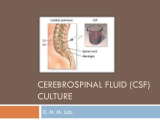
LUMBAR PUNCTURE A lumbar puncture also called a spinal tap is a procedure where a sample of cerebrospinal fluid is taken for examination. CSF is mainly used to diagnose meningitis [an infection of the meninges]. It is also used to diagnose some other conditions of the brain and spinal cord.
PRECAUTIONS FOR LUMBAR PUNCTURE • Asked to sign a consent form • Ask about taking any medicines • Are allergic to any medicines • Have / had any bleeding problems • Ask about medications such as aspirin or warfarin • Ask the female patient might be pregnant • Empty the bladder before the procedure
LUMBAR PUNCTURE 1. Material for sterile technique [gloves and mask are necessary]2. Spinal Needle, 20 and 22-gauge3. Manometer4. Three-way stopcock5. Sterile drapes6. 1% lidocaine without epinephrine in a 5-cc syringe with a 22 and 25-gauge needles7. Material for skin sterilization8. Adhesive dressing9. Sponges - 10 X 10 cm
LUMBAR PUNCTURE [Complications] Post lumbar puncture headache occurs in 10% to 30% of patients within 1 to 3 days and lasts 2 to 7 days. The pain is relieved by lying flat. Treatment consists of bed rest and fluid with simple analgesics.
LUMBAR PUNCTURE [Complications] Headache following a lumbar puncture is a common and often debilitating syndrome. Continued leakage of cerebrospinal fluid from a puncture site decreases intracranial pressure, which leads to traction on pain-sensitive intracranial structures. The headache is characteristically postural, often associated with nausea and optic, vestibular, or otic symptoms. Although usually self-limited after a few days, severe postural pain can incapacitate the patient. Management is mainly symptomatic, but definitive treatment with the epidural blood patching technique is safe and effective when done by an expert operator.
LUMBAR PUNCTURE Patient usually lie on a bed on side with knees pulled up against the chest. It may also done with sitting up and leaning forward on some pillows. Sterilize the area. push a needle through the skin and tissues between two vertebra into the space around the spinal cord which is filled with CSF. CSF leaks back through the needle and is collected in a sterile container. As soon as the required amount of fluid is collected the needle is taken out and a plaster is put over the site of needle entry.
LUMBAR PUNCTURE Sent the sample to lab to be examined under the microscope to look for bacteria. It is also 'cultured' for any bacterial growth The fluid can also be tested for protein, sugar and other chemicals if necessary. Sometimes also measure the pressure of the fluid. This is done by attaching a special tube to the needle which can measure the pressure of the fluid coming out.
LUMBAR PUNCTURE Place the patient in the lateral decubitus position lying on the edge of the bed and facing away from operator. Place the patient in a knee-chest position with the neck flexed. The patient's head should rest on a pillow, so that the entire cranio-spinal axis is parallel to the bed. Sitting position is the second choice because there may be a greater risk of herniation and CSF pressure cannot be measured
LUMBAR PUNCTURE Find the posterior iliac crest and palpate the L4 spinous process, and mark the spot with a fingernail. Prepare the skin by starting at the puncture site. Anesthetize the skin using the 1% lidocaine in the 5 mL syringe with the 25-gauge needle. Change to 22-gauge needle before anesthetizing between the spinous process. Insert in the midline with the needle parallel to the floor and the point directed toward the patient's umbilicus
LUMBAR PUNCTURE Advance slowly about 2 cm or until a "pop'' (piercing a membrane of the dura) is heard. Then withdraw the stylet in every 2- to 3-mm advance of the needle to check for CSF return. If the needle meets the bone or if blood returns (hitting the venous plexus anterior to the spinal canal), withdraw to the skin and redirect the needle. If CSF return cannot be obtained, try one disk space down