Download
1 / 39
390 likes | 399 Vues
Intracranial Catastrophe. Clinical Associate Professor Andrew Bezzina September 2013. Disclosures. Dr. Andrew J. Bezzina Not clever enough to have anything to disclose. Meet Uncle Frank. 72 year old male collapsed at home.

E N D
Intracranial Catastrophe Clinical Associate Professor Andrew Bezzina September 2013
Disclosures • Dr. Andrew J. Bezzina • Not clever enough to have anything to disclose.
Meet Uncle Frank. • 72 year old male collapsed at home. • Complains of a fronto-temporal headache and a left hemiparesis with onset at time of the collapse. • A and B – Normal • C - PR 92, BP 184/96 • D - GCS 14 • Left hemiparesis, distal>proximal and 3/5 power. • Decreased sensation on left to all modalities
Strategy? • Time critical intervention? • How do we predict progression? • Strategies that will improve his prognosis? • We know what we do but do we know what we do works?
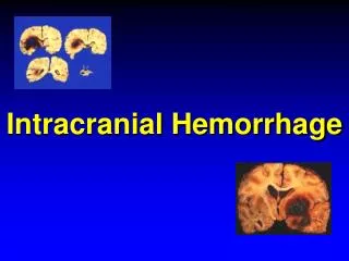
Intracerebral Bleeds –Scope of The Problem • 10 – 20/100,000 per year • 10% to 15% of new strokes that occur every year in Europe, US and Australia • 20 -30% in Asia. • Thirty-day mortality • 30% to 50% • 20% independence at 6 months. • 1 year mortality 75% • Traditionally chronic hypertension the cause in 80-90% BUT more recent data only about 50 - 65% Lancet Neurol 2010; 9: 167–76.
What’s changing? • Better BP control • Increasing age of population • More use of anticoagulants • Improved imaging techniques = detection of smaller and more subtle bleeds.
What’s the nature of the problem? • Primary Intracerebral Haemorrhage • spontaneous rupture of small vessels • 78 to 88% of all cases • Secondary Intracerebral Haemorrhage • other than small vessel rupture (e.g., aneurysm, AV malformation, haemorrhagic transformation of ischaemic stroke, neoplasms) • 12 to 22% of all ICH
Pathophysiology • Haematoma • Haematoma Expansion • Ischaemia ? • Secondary injury • Cascade initiated by the primary injury (e.g. mass effect and physical disruption) • Physiological response to the haematoma (e.g. Inflammation – inflam cells/ complement) • Toxic clot components – Thrombin /Hb and Iron Free radicals
Can we predict progress? • We’ve got the CT result and diagnosis can we tell who is likely to get worse?
Early Clinical Picture • > 20% will have decrease in GCS of 2 points between pre-hospital assessment and the initial evaluation in ED • If pre-hospital deterioration - GCS score decreases by an average of 6 points and the mortality rate is 75%. • Within first hour of presentation to a hospital, 15% of patients demonstrate a decrease in the GCS score of 2 points. • No surprises • Sicker patients = faster deterioration = higher mortality • Underscores the need for aggressive early management.
Predictors in ED • Retrospective Cohort Study • 619 patients SICH (excluded patients on Warfarin), 22.6% had ED neurologic deterioration. • Independent predictors - • regular antiplatelet use, • ictus to arrival time < 3 hours, • initial body temperature > 37.5C, • associated intraventricular haemorrhage • midline shift > 2 mm on CT. • associated with 1-week mortality, 30-day mortality, and poor neurologic outcome on discharge • ECG changes and troponin leaks no effect on short-term prognosis Ju-Sing Fan, MD et al. acad. Emerg. Med. 2012; 19:133–138
Location, Location, Location • Outcome and potential treatment. • Pontine haemorrhages highest mortality • Superficial haemorrhages might be more amenable to surgery. • Intraventricular haemorrhages - predict a poor outcome. • 30-day mortality rates vary with location: • 44% for deep ICH, • 46% for lobar ICH • 60% for brainstem ICH • 34% for cerebellar ICH .Neurology 2006;66:1182–1186.
Haematoma Volume • Initial volume - strongest predictor of 30 - day mortality, with 96% sensitivity and 98% specificity • 10% enlargement = 5% increased death • 10% enlargement = 16% increase in the likelihood of 1 point worsening on the modified Rankin
Haematoma Expansion • 73% some degree of expansion. • Roughly 1/3 will have > 1/3 expansion • Occurs early - 75% first 6 hours. Rest within 24 hours. • Risk ∝ Initial size • Other risk factors for expansion – • early presentation • warfarin use • contrast extravasation on CT angiography (CTA) .
Anticoagulants • Major Trials - rates of major haemorrhage on warfarin typically 1% to 3% per person-year • Observational studies - higher outside of a clinical trial setting e.g. 13.1% per person year (4.1% under age 80) • No large studies offering real-world, population-based estimates of haemorrhage rates among patients taking warfarin
High Risk Group on OAC • Long-term anticoagulation = 7 – 10 fold increase in the risk of ICH • Increased risk of ICH in patients on long term anticoagulation if • older age • hypertension • previous ischaemic stroke • leukoariosis • INR > 3.5 (most still occur below that level) • Antiplatelet use = OAC assoc ICH.
Antiplatelet Agents and Haematoma Expansion • Antiplatelet use was not found to be associated with haematoma expansion in one neuroprotective study However • Naidech and colleagues reported worse 3-month outcomes in patients with decreased platelet function. • A systematic review suggests that on balance previous antiplatelet use associated with increased mortality Stroke 2009;40(7):2398Y2401.
CT “SPOT” • Spot Sign – “spot-like and/or serpiginous foci of enhancement, within the margin of a parenchymal hematoma without connection to outside vessels. Greater than 1.5 mm in maximal dimension and a Hounsfield unit density > background hematoma density”.
Spot Sign • Identified in 19% of 367 CT angiograms in one study. • Spot sign positive = 69% risk of expansion. • Scoring system (based on the Hounsfield units, the number of spots, and the size of the initial haemorrhage) independent predictor of in-hospital mortality and 3-month outcome. • Guidelines - CT angiography a Class IIb recommendation for identification of hematoma expansion.
Implications of the Spot Sign • Prevalence of spot sign decreases as time from onset to CTA increases • Presence remains predictive of haematoma expansion independent of time from onset to CTA . • Several validating studies - predictive of mortality and poor outcome. • Secondary ICH spot sign also predictive of in-hospital mortality. • vascular and nonvascular mimics lower its sensitivity and specificity.
Can we do anything? • NO proven (in phase 3 trials) medical or surgical treatment for intracerebral haemorrhage exists • Surgical decompression - widely accepted as potentially life-saving in cerebellar haemorrhage. • Treatment in a neurology or neurosurgery intensive care unit may well improve outcomes.
Basics • ABC – • keep them alive • avoid secondary injury • Head Elevation • Early transfer/retrieval
Stop the Bleeding – FFP? Systematic Review • “In summary, .........at an intuitive level it seems to be an appropriate product to replace or supplement different constituents of plasma in patients. ......... Has not been subjected to the clinical research scrutiny that should be required to demonstrate effectiveness.” . British Journal of Haematology, 2004, 126, 139–152
Reversal of Anticoagulation • Correction of the INR to less than 1.4 within 2 hours -associated with decreased haematoma expansion (DOE) • Likelihood of INR correction at 24 hours was linked to time to FFP administration in 1 study, 17% of patients still did not have an INR < 1.4 at this time. • Prospective multicenter registry of patients with aaICH - found PCC therapy reversed anticoagulation in 71.8% of patients within 1 hour of treatment. • Significant haematoma expansion still occurred in 47% and the in-hospital mortality rate was 44% • Thrombotic events occurred within 30 days of treatment, including strokes, AMI, and venous thrombosis. 2% rate over 7 days. CanPro Registry. Stroke. 2011;42:e586–e629.
Other haemostatics - • 5 phase II RCTs and 1phase III RCT, involving 1398 adults aged 18 years or over, within four hours of ICH onset: • 423 received placebo and 975 received haemostatic drugs (2 received epsilon-aminocaproic acid (EACA) and 973 received recombinant activated factor VII (rFVIIa)). • Haemostatic drugs did not significantly reduce 90-day case fatality after ICH (risk ratio (RR) 0.85 ;CI 0.58 to 1.25), • rFVIIa did not significantly reduce death or dependence on the modified Rankin Scale (grades 4 to 6) within 90 days of ICH (RR 0.91; CI 0.72 to 1.15). • rFVIIa trend to thrombo-embolic serious adverse events (RR 1.37, 95% CI 0.74 to 2.55)
Other haemostatics • Systematic review – suggested previous use of anti-platelet therapy is associated with increased mortality. • Can anti-platelet therapy be “reversed”? • Transfusion of fresh platelets - value unclear and is considered investigational. • Under investigation in the Platelet Transfusion in Cerebral Haemorrhage (PATCH) trial (The Netherlands National Trial Register No. NTR1303)
High BP – what’s the harm? A systematic review and a recent large multisite study in China • Systolic BP > 140 to 150 mm Hg within 12 hours of ICH associated with more than double the risk of subsequent death or dependency. • Cause? Effect?
INTERACT Conclusions • intensive lowering of blood pressure did not result in a significant reduction in the rate of the primary outcome of death or severe disability. • The ordinal analysis showed significantly lower modified Rankin scores with intensive treatment (odds ratio for greater disability, 0.87; 95% CI, 0.77 to 1.00; P = 0.04). Secondary outcome.
Anticonvulsants • Incidence of clinical seizures – reports up to 17% within the first few weeks • Continuous EEG monitoring - seizures in almost one third of patients despite prophylactic anticonvulsants. • 2 observational studies have reported worse outcomes in patients prophylactically treated with phenytoin after ICH. • One large, single-centre study, prophylactic antiepileptic drugs did significantly reduce the number of clinical seizures after lobar ICH However • Prospective and population-based studies, clinical seizures not associated with worse neurological outcome or mortality.
Subclinical seizures (EEG only) • A recent analysis from placebo arm of an ICH neuroprotectant study • Patients who received antiepileptic drugs (primarily phenytoin) without a documented seizure were significantly more likely to be dead or disabled at 90 days, after adjusting for other established predictors of ICH outcome Messe´ SR et al. CHANT investigators. Prophylactic antiepileptic drug use is associated with poor outcome following ICH. Neurocrit Care. 2009;11: 38–44.
Osmotic Treatment Randomised trials • No significant benefit of bolus therapy with mannitol on cerebral blood flow, mortality and functional outcomes in ICH • Evidence from smaller trials that mannitol can help to buy time for 24–48 h, before more definitive therapy can be instituted. • Review of HTS versus Mannitol • Control of intracranial pressure 93% with hypertonic sodium solution 78% with mannitol (pooled RR hypertonic sodium 1.16 95% CI 1.00-1.33).
Surgery Current practice surgical intervention if - • Superficial lobar haemorrhage, GCS 9–12, volume 20–80 ml, and ⁄ or deteriorating. • Clot volume between 20 and 80 mls. • Worsening neurological status. • Relatively young patients. • Haemorrhage causing midline shift ⁄ raised ICP. • Cerebellar haematomas > 3 cms &⁄ or causing hydrocephalus. • Putaminal haematoma with GCS 9–12, volume 20– 60 ml and deteriorating.
Surgery?? • Most surgical trials - supratentorial ICH - No benefit. • Surgical Trial in IntracerebralHemorrhage (STICH) - No benefit. • This trial randomized over1000 patients. surgery versus best medical practice based on the theory of equipoise (Patients were eligible for the study only if the responsible physician was uncertain as to benefits of either treatment). • Six month mortality, Glasgow Outcome Scale scores, modified Rankin Scale scores, and Barthel Index scores nearly identical.
CerebellarHaemorrhage • Several nonrandomized studies • Beneficial outcomes for cerebellar hematomas when surgically evacuated compared to medical treatment only if : • a diameter exceeding 3 cm, • causing hydrocephalus or brainstem compression,
Stereotactic Clot Removal Stereotactic Treatment of Intracerebral Hematoma by Means of a Plasminogen Activator (SICHPA) trial, • significant volume reduction in the treatment arm of 10 to 20% within 72 hours of symptom onset. • No significant effect on 6-month functional outcome or mortality 2 other trials (one nonrandomized and one randomized found stereotactic aspiration of deep ICH • safe and associated with early improvement of National Institute of Health Stroke Scale (NIHSS)scores and improvement of long-term functional outcome measured by modified Rankin Scale. Early Days
New Medical Therapies • Desferoxamine • Anti-inflammatories • Celecoxib • Pioglitazone • Statins • Albumin
So back to Frank… • ABC • If oral anticoagulation consider reversal using PCC +/- FFP • If anti-platelet agent probably no role for platelet transfusion • Leave BP alone BUT if you’re tempted be very careful. • No anticonvulsants. • Mannitol if you have to • Discuss early with neurosurgeons