Download
1 / 56
580 likes | 921 Vues
A 58-year-old woman with recurrent RUQ pain. Julie Fagan, M.D. Primary Care Conference 7 July 2004. Learning Objectives. Recognize biliary sources of abdominal pain List differential diagnosis of RUQ pain Describe diagnostic algorithm for evaluating RUQ pain

E N D
A 58-year-old woman with recurrent RUQ pain Julie Fagan, M.D. Primary Care Conference 7 July 2004
Learning Objectives • Recognize biliary sources of abdominal pain • List differential diagnosis of RUQ pain • Describe diagnostic algorithm for evaluating RUQ pain • Evaluate evidence for treatment of various causes of RUQ pain
COI Disclaimer No financial support was given for this presentation
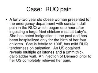
Case Presentation • 58 y/o WF with recurrent abdominal pain for many yrs • 11/97: recurrent epigastric/upper abdominal pain relieved by H2 blockers, omeprazole • FHx: + for GERD, metastatic adenoCA of unknown primary, likely GI • SHx: married, 2 children, technology instructor, no tobacco, EtOH, NSAIDs • PMHx: obesity (fen/phen tx), no surgery
Case Presentation • Meds: omeprazole 20 mg/day, MVI • ROS: negative for wt loss; positive for occasional diarrhea, nausea, radiation to back (flank) • Exam: obese, no jaundice, mild epigastric tenderness, no masses or peritoneal signs, otherwise normal
Case Presentation • Labs: CBC, ESR, LFTs, amylase, lipase WNL • GB u/s in past had showed GB polyps, no gallstones • Pt was referred to GI, who diagnosed her with IBS, GERD and recommended a barium enema, which was normal
Case • 1/6/99: attack of sharp RUQ pain radiating to back, shoulder, lasted 20 minutes. Took extra Gas-Ex, famotidine • 5/99: EGD showed nonobstructing distal esophageal stricture, diaphragmatic hernia • Seen periodically in next 5 yrs with dyspepsia, GERD sx, RUQ pain
Case • 6/04: complained of most severe episode ever of RUQ pain radiating to back • Recent intentional 60-lb weight loss • Self- discontinued omeprazole, no more GERD sx • Pain lasted several hours, triggered by eating cheese
Case • Exam: Weight 174, no sclericterus. 140/84, 84. Afebrile • Abdomen: no masses, tenderness, hepatosplenomegaly • Labs: again, all nl (CBC, chem 7, LFTs, amylase, lipase
Differential Diagnosis • Recurrent pancreatitis • Acalculous biliary pain (ABP) • Malignancy of gallbladder, pancreas, CBD • Dyspepsia • Gastritis or PUD • Non-ulcer dyspepsia
Differential Diagnosis • IBS/functional abdominal pain • But no alterations in bowel function • Dysfunction of the sphincter of Oddi • R sided pyelonephritis • Hepatic or subphrenic abscess
How to Proceed Next? • HIDA (hepatobiliary) scan 6/04: • No ejection fraction response to cholecystokinin after 30 minutes • Consistent with biliary dyskinesis
Sphincter of Oddi Dysfunction (SOD) • Definition: a functional disturbance of the sphincter of Oddi (SO) leading to intermittent biliary obstruction • Other terms used: • Papillary stenosis, sclerosing papillitis, biliary spasm, stenosis of the SO, biliary dyskinesia, post-cholecystectomy syndrome
Function of the Sphincter of Oddi • To maintain a low-pressure system within the hepatic ducts • Allows hepatic secretion to proceed irrespective of bile flow rate
Symptoms of SOD • 2 distinct syndromes • Biliary pain without other causes • Frequently seen after cholecystectomy • Surgery may unmask SOD, since gallbladder can decompress biliary system during sphincter spasm • Idiopathic recurrent pancreatitis • May explain many cases of post-ERCP pancreatitis
Symptoms • Pancreatitis: unsure whether cause or effect • 22% of pts with known cause of pancreatitis (e.g. alcohol, gallstones) had elevated SO pressures • SOD found in up to 87% of patients with chronic pancreatitis in another study
Diagnosis • Gold standard: SO manometry during ERCP • Less invasive tests: • Dilation of CBD (5-10 mm) • Occurs in 16% of asymptomatic pts after cholecystectomy • Increases with age • Provocative testing
Diagnosis • Provocative tests to increase bile flow • Fatty meal ultrasonography • Cholecystokinin • In normal subjects, bile duct diameter should stay the same or decrease • An increase of > 2 mm considered abnl • Hepatobiliary scintigraphy
HIDA scans • HIDA is an iminodiacetic acid derivative, which is given by intravenous injection, taken up by hepatocytes, and excreted into bile with concentration in the gallbladder. Failure to opacify the gallbladder is the most sensitive and specific finding
Hepatobiliary Scintigraphy • HDTT (transit time from hepatic hilum to duodenum is measured in post-cholecystectomy patients • Gallbladder ejection fraction (amount of contrast that leaves the gallbladder after a CCK injection for patients with suspected acalculous biliary pain
Accuracy of HIDA • Varying results in post-cholecystectomy patients when compared with SO manometry • Sensitivity: 13% • Specificity: 95% • Positive predictive value: 50% • Negative predictive value: 74% Craig et al, Gut 2003
Classification of SOD • Methods of predicting SOD and its response to treatment • Milwaukee classification (Hogan & Geenen) is most widely used • Based on clinical presentation, lab results, ERCP findings • Divides patients with biliary pain into 3 groups
Milwaukee Classification of SOD • Type I: all 4 criteria • Type II: biliary-type pain and 1 or 2 of the other criteria • Type III: biliary pain only, with no other objective abnormalities
Probability of SOD According to Classification • Type I: 65-95% • Type II: 50-63% • Type III: 12-28%
Pathophysiology • SO is a small sphincter that regulates the flow of bile and pancreatic enzymes into duodenum and prevents reflux of duodenal contents into ducts • SOD thought to cause pain by impeding flow of bile and pancreatic juice, with ductal hypertensiondistention and inflammation
Pathophysiology of SOD • Type 1 patients more likely to have structural abnormalities (papillary stenosis) • Conversely, Type III patients more likely to have dysmotility, high basal pressure, rapid contraction frequency, paradoxical response to CCK, excessive retrograde contractions • High concordance of IBS
Clinical Presentation • Classic patient: female 20-50 with history of recurrent abdominal pain • RUQ or epigastric • Disabling • Lasts 30 minutes-several hours • May radiate to back or shoulder • May have N & V • May occur years after cholecystectomy
Clinical Presentation • Some pts have continual pain not relieved by surgery • Jaundice, fever, chills are rare
Physical Exam • Paucity of abnormal findings is the rule • Most common: mild, nonspecific abdominal tenderness • Pain usually not relieved by H2 blockers, PPI’s, antacids, IBS medications • Some pts present with typical pancreatic pain, recurrent pancreatitis
Diagnosis • LFTs (especially at time of biliary pain • AST, alk phos, bili > 2 X upper limit of nl • Significant increase in amylase or lipase closely temporally related to pain episodes • Endoscopy to r/o UGI disease • Ultrasound to r/o gallstones, check CBD
Diagnosis • Ultrasound (cont) • A CBD diameter > 6 mm may indicate increased resistance through the SO • But found in 34% of asymptomatic cholecystectomized patients • Fatty-meal or CCK-enhanced ultrasound • Patients without SOD should have contraction of CBD, or no change
Diagnosis: Nardi Test • Morphine-prostigmine administered simultaneously induce SO spasm and pancreatic exocrine secretion • Rarely used: low sensitivity and specificity • Poor correlation with outcome after sphincterotomy • Replaced by other evaluations today
Diagnosis: Hepatobiliary Scintigraphy • Quantitation of bile flow through biliary tract • False positives due to stones, tumors, sphincter disease • Precise criteria for positive test controversial: time to peak, half-time of excretion, duodenal appearance time, hilum-to-duodenum transit time • Most studies not correlated with manometry
MRCP • New tool with high sensitivity and specificity in distinguishing benign from malignant lesions (Lee et al Radiology 1997) • Artifacts seen in presence of metal clips • Major limitation: unable to offer therapeutic options
Endoscopic Ultrasound • Most helpful for detecting microlithiasis (sludge) in ampullary region • Compared to MRI: 97% sensitivity and 88% specificity in diagnosing extrahepatic biliary obstruction • Fewer complications than ERCP, but invasive procedure often performed under general anesthesia by specialists
ERCP • Essential to r/o stones, tumors, or other obstructing processes of biliary tree • Definitive supine drainage times of biliary tree not well defined yet, but post-cholecystectomy biliary tree that fails to drain by 45 minutes generally considered abnormal
Sphincter of Oddi Manometry • Gold standard to diagnose SOD and stratify therapy • Helpful selecting pts more likely to benefit from sphincterotomy • Baseline sphincter pressure most helpful parameter • Most important finding for SOD: basal pressure > 40 mm Hg
SO Manometry • Frequency of abnormal pressure 7-65% overall • Range: 28-60% in Type II biliary pts • 7-55% among Type III biliary pts • Explanations for variance • Selection of pts with biliary-type vs nonspecific abdominal pain increases yield of abnormal SOM
SO Manometry • Patients with SOD divided into 2 groups • Structural alterations of SO zone (stenosis) • Functional abnormalities (dyskinesia)
Sphincter of Oddi Stenosis • Basal SO pressure abnormally elevated (> 40 mm Hg)
Sphincter of Oddi Dyskinesia • Pts may also have elevated basal SO pressures • Administration of amyl nitrate or glucagon (to relax smooth muscles) dramatically decreases pressure • Smooth muscle relaxants do not reduce pressure in pts with SO stenosis
Other Features of SO Dyskinesia • Rapid SO contraction frequency (> 7/min) • Excessive retrograde phasic contractions (>50%) • Paradoxical basal pressure increase after CCK-8 administration
Limitations of SOM • Increased risk of pancreatitis (17% in some series) • Compared to post-ERCP rate of 5% • Other studies suggest that SOD, not manometry, increases pancreatitis risk • Technique, equipment, sedation, observer experience affect results
Treatment • Pharmacologic • Calcium-channel blockers • Nifedipine • Nitrates (case reports only) • Electroacupuncture • Endoscopic sphincterotomy • Botox • Surgery
Nifedipine Effect on SOD • Conflicting results • Sublingual nifedipine (10-20 mg) had no effect on SO pressure (Guelrud et al, Gastroenterology 95:1050) • Double-blind crossover trial showed improvement in biliary pain in 21/28 patients with SOD (Khuroo et al, Br. J Clin. Pharmacol. 33:477)
Electroacupuncture • One report from Korea (Lee et al Gastrointestinal Endoscopy 53:211)showed significant decreased SO pressure, amplitude and frequency of phasic contractions with stimulation of R lateral tibia • Clinical relevance uncertain
Conclusions: Medical Therapy • Few studies use SOM as gold standard • Symptoms recur after therapy stopped • Most recommend medical therapy to screen Type I and II pts for sphincterotomy benefit • Most recommend a trial of medical therapy for type III patients • Low-dose antidepressants, antispasmodics
Therapy: ES • Currently the recommended treatment for most patients • Surgery reserved for restenosis or when endoscopic therapy not available or feasible • Pain relief for Type I and II pts with ES is 90-95%