Download
1 / 31
410 likes | 1.08k Vues
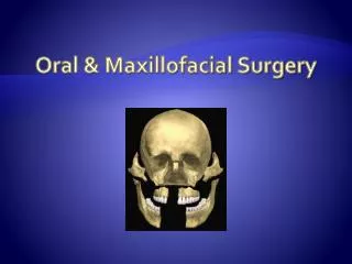
Oral & Maxillofacial Surgery. Oral & Maxillofacial Surgery. Oral and maxillofacial surgery is surgery to correct a wide spectrum of diseases, injuries and defects in the head, neck, face, jaws and the hard and soft tissues of the oral and maxillofacial region.

E N D
Oral & Maxillofacial Surgery • Oral and maxillofacial surgery is surgery to correct a wide spectrum of diseases, injuries and defects in the head, neck, face, jaws and the hard and soft tissues of the oral and maxillofacial region. • An oral and maxillofacial surgeon is a regional specialist surgeon treating the entire craniomaxillofacial complex: anatomical area of the mouth, jaws, face, skull, as well as associated structures.
-The Extreme-OM surgery plus Plastics plus ENT, plus... These recently updated photos obtained May 5, 2009 from the Cleveland Clinic shows Connie Culp after an injury to her face and as she appears today. Five years ago, a shotgun blast left a ghastly hole where the middle of her face had been. Five months ago, she received a new face from a deceased woman.
A multi-disciplinary team of doctors and surgeons at Cleveland Clinic performed the first near-total face transplant in the United States. In a 22-hour procedure, surgeons transplanted 80 percent of a woman's face who suffered severe facial trauma � essentially replacing her entire face, except for her upper eyelids, forehead, lower lip and chin.
Alveolar process - alveolar ridge: a ridge that forms the borders of the upper and lower jaws and contains the sockets of the teeth Arthroscopy Calvarial - skull Condyle - A rounded enlargement or process possessing an artculating surface. Coronal flap Craniosynostosis - Premature closing of joints or sutures in the skull. Dentition - teeth Glenoidfossa- jaw joint Inferior alveolar nerve -enters the mandible on the deep surface of the ramus, providing sensation to the teeth Gnath-jaw Labia - lip Malar bone - cheek Malocclusion Maxillofacial - relating to the lower half of the face Meniscus - cartilage that works to absorb weight and provide stability Mouth prop Mental Nerve - nerve which provides sensation to the anterior aspects of the chin and lower lip as well as the buccalgingivae of the mandibular anterior teeth and the premolars. Orbicular- circular in outline Terms
Terms • Osteotomy - operation in which the bone is cut through • Ramus - the posterior part of the mandible that is more or less vertical • Reduction • Sagittal -[sagitt = arrow] Divides the body or structure into mirror images of right & left sides. • Symphysis - the point of junction of two bones as in the two parts of the lower jaw in front: the tip of the chin • TMJ - Tempero-mandibular joint, the two joints that connect the jaw to the skull.
Oral (Buccal) Cavity • Lips • Teeth • Palate • Cheeks • Tongue
Lips (labia) • Thermal receptors for protection from burns • Muscles aiding in expressions, food retention, and mastication • Space between lips extending to the cheeks is the vestibule (an entrance)
Deciduous (primary) Appear = 6 mos. Continue to = 4 yrs For each jaw: 2 central incisors 2 lateral incisors 2 cuspids or canines 4 molars 20 total Loss begins = 6 yrs Permanent (secondary) For each jaw: 2 central incisors 2 lateral incisors 2 cuspids or canines 4 bicuspids 6 molars 3rd molars or wisdom teeth appear = 17-25 yrs Teeth (Dentitions)Rest in alveolar processes
Teeth • Speech • Mastication
1. Crown is above/outside gumline Enamel covering of crown Hardest part Doesn’t reproduce Degenerates with age and injury Dentin Majority of tooth Harder than bone Encases pulp Pulp Blood vessels Nerves Connective tissue Teeth(3 Regions)
Teeth(3 regions) • Neck • Junction of crown and root
Teeth(3 regions) • Root • Held by periodontal ligament • Connects tooth to alveolar processes
Palate • Roof of the mouth • Anterior portion is hard palate • Posterior portion is soft palate • Uvula is the most posterior part of the soft palate • Soft palate separates mouth from nasopharynx • Soft palate rises with swallowing to prevent food going into nasal cavity
Cheeks • Lateral walls of oral cavity • Consist of major muscles for mastication
Tongue • Chemoreceptors for taste • Attached to floor of buccal cavity by lingual frenulum • Function in speech, propelling food through oral cavity and swallowing
Axial Skeleton Along or connecting to the midline of the body Skull Vertebral column Sternum Rib Cage About 80 bones total Appendicular Skeleton Everything hanging off the axial skeleton Skeletal System
Frontal Temporal Occipital Parietal Sphenoid Ethmoid Maxilla Zygomatic Mandible Lacrimal Hyoid Skull
Skull • Frontal bone • Protection • Twice as thick as others • Supraorbital foramen allow for passage of blood vessels and nerves servicing the face and head
Skull • Temporal Bone • Protection • External auditory canal allows for sound to enter skull • Openings at base for carotid arteries and jugular veins called carotid foramen and jugular foramen • Mastoid process where sternocleidomastoid muscle attaches
Skull • Occipital bone • Forms posterior base of skull • Large hole at base is foramen magnum where spine enters skull • Occipital condyles are connections between skull and vertebrae • C-1 vertebrae most superior is the atlas which supports the skull/head • C-2 is the axis which allows for movement of the head
Skull • Sphenoid bone - butterfly-shaped bone at the base of the skull • Center of base of skull • Keystone of the skull (ties a lot of other bones together • Sella turcica is where the pituitary gland sits - a saddle-shaped depression in the sphenoid bone at the base of the human skull.
Skull • Ethmoid bone • Forms part of nasal cavities • Olfactory foramen open into nasal cavity • Penetrates into frontal bone - cribiform bone • Nerve ending in cribiform plate where sense of smell delivered and received
Skull • Maxilla • Attachment for upper teeth (alveolar processes) • Infraorbital foramen for vessels and nerves • Attachment for zygomatic bones/cheek bones
Skull • Mandible • Lower jaw • Largest and only moving bone in face • Attachment for lower teeth (alveolar processes) • Articulates with glenoid fossa of each temporal bone = TMJ (temporomandibular joint)
Skull • Lacrimal bone • Smallest bone in face – very thin • Small foramen become nasolacrimal ducts where tears drain • Tears come from lacrimal gland in eyebrows (keep eyes and nares moist)
Skull • Hyoid bone • Only bone in body that does NOT connect directly to another bone • Held in place by ligaments • Holds tongue in place (tongue for swallowing and mastication)
Orbital Bones (* =‘s weakest bones) • Frontal • Lacrimal • Ethmoid-* • Maxilla- * • Zygomatic • Sphenoid • Palatine
Facial Muscles • Expressions • Mastication • Speaking