Download
1 / 22
310 likes | 925 Vues
Osteomyelitis. Dr/Wael H. Mansy, M.D. Assistant Professor King Saud University. Objectives of this session:. Familiarize the audience with major types of Osteomyelitis, relationship to the age of the host, mechanism of infection, bacteriology of the disease. Clues that help in the diagnosis

E N D
Osteomyelitis Dr/Wael H. Mansy, M.D. Assistant Professor King Saud University
Objectives of this session: • Familiarize the audience with major types of Osteomyelitis, relationship to the age of the host, mechanism of infection, bacteriology of the disease. • Clues that help in the diagnosis • Overview of the management of Osteomyelitis.
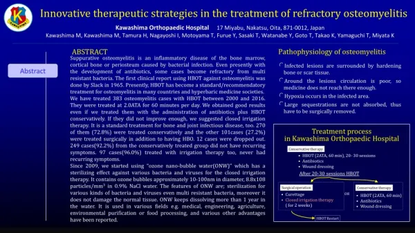
Osteomyelitis: Definition • Infection of the bone and bone marrow (osteo, myelitis) • Mostly bacterial, can be fungal
Epidemiology • Pre-antibiotic era had 25% mortality • Significant morbidity/disability worldwide due to lack of access to care • Leading cause for amputations in the US • Significant cause of pediatric disability worldwide.
Prevalence: • Children 1: 5000 • Sickle cell patients 3.6: 1000 • Post puncture wound to foot 16% • Neonates 1: 1000 • Post puncture wound to foot in diabetics 30 – 40% • Higher in developing countries
Osteomyelitis • Usually subdivided clinically into: • Pediatric • Adult • Hematogenous vs. Direct spread • Special cases of Intravenous Drug Abusers (IVDA) and Sickle cell Anemia.
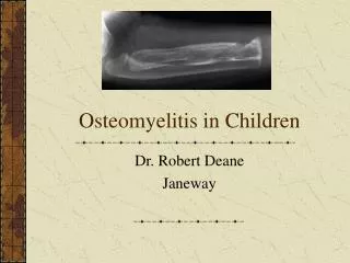
Pediatric Osteomyelitis • Hematogenous spread affecting the long bones. • Usual sites are the long bones: tibia, humerus, femur. • Some to Spine: direct contact (TB)
Why the long bones? • Non-anastomosing capillary ends of nutrient arteries form sharp loops under the growth plates and enter large venous sinusoids where the blood flow is slow and turbulent, trapping the organisms.
Usual causative organisms: Pedi. Osteo. • Staphylococcal aureus • Streptococcus suppurefaticus. • Hemophilus. influenza. • Sickle cell disease: Long bone osteomyelitis often due to salmonella.
Adult Osteomyelitis • Most Cases: Direct extension of infection to the bone from a skin ulceration, leading cause of amputations • Direct inoculation to the bone from an open/contaminated fracture. • Hematogenous from IVDA
Adult Osteomyelitis • IVDA: Hematogenous site more likely to be spine or pelvis only occasionally to the long bones.
Adult Osteomyelitis • Most common: Foot ulcer extending into the bony structures. • Neuropathic foot ulcer • Mixed infection is common with s.aureus, Gram negatives, some strep. • Open fractures • Infected prostheses • Foot injuries
Management: Admit • OrthoSurg consultation • Closed needle biopsy/drainage • C/S obtained • Started on I.V. vancomycin empirically • Switched to oxacillin after C/S grew meth. Sensitive staph. A.
Hosp. Course • Over 6 to 8 days on I.V. antibiotic therapy, patient became afebrile, leg tenderness subsided, less pain w/ ambulation. • On 9th day patient switched to oral penicillin, sent home to complete 6 weeks of therapy. • Full recovery when seen for follow up visit in clinic.
Osteomyelitits • Follow up: can consider repeat ESR have it return to normal level. • Follow up films, radiologic recovery slower than clinical recovery