Download
1 / 36
390 likes | 965 Vues
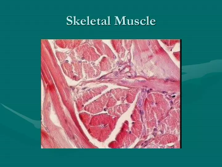
Skeletal Muscle. Skeletal Muscle Anatomy. Figure 12-3a-2: ANATOMY SUMMARY: Skeletal Muscle. Figure 12-4: T-tubules and the sarcoplasmic reticulum. Myofibrils: Site of Contraction. Figure 12-3c-f: ANATOMY SUMMARY: Skeletal Muscle. Smooth muscle fascicles !. Smooth Muscle.

E N D
Skeletal Muscle Anatomy Figure 12-3a-2: ANATOMY SUMMARY: Skeletal Muscle
Myofibrils: Site of Contraction Figure 12-3c-f: ANATOMY SUMMARY: Skeletal Muscle
Smooth Muscle The smooth muscle looks smooth and works automatically. It can be found in the intestines, lungs, and bladder.
Cardiac Muscle Cardiac muscle is located only in the heart. It has a striated appearance and is involuntarily controlled. Cardiac muscle also has a feature that is foreign to the other muscle types: intercalated discs.
This muscle is striped and branched (like a tree!) It keeps your heart beating automatically. Where is it found? Intercalated Disc
VII. Interactions of Skeletal Muscles in the Body A. Attachment Points (could be one or multiple): 1. Origin- point of attachment on bone; does not move during contraction 2. Insertion- point of attachment; does move during contraction
Muscles work in groups- some contract and some relax and some assist. • Four functional classifications • Prime Movers • Antagonist • Synergist • Fixators
Prime Mover • Agonist • Provides the major force for producing a specific movement • For elbow flexion: biceps brachii
Antagonists • Muscles that oppose, or reverse, a particular movement • When the prime mover is active, the antagonist muscles are often stretched and may be relaxed • Ex. Triceps brachii- rest during flex • Prime movers and its antagonist are located on opposite sides of the joints • Flexion of the arm by the biceps brachii muscle of the arm is antagonized by the triceps brachii, the prime mover for extending the forearm
Synergists • Help the prime movers by • Adding a little extra force • Reducing undesirable or unnecessary movements that might occur • Ex. Brachioradialis- assists in flex They act as stabilizers: When muscles cross two or more joints it’s contraction causes movement of all the spanned joints so these muscles act to stabilize the joints
Fixators • Synergist that immobilize a bone • Fixator- joint stabilizers; maintain posture/balance • Ex. Deltoid- maintains shoulder joint stability • Scapula is held to the axial skeleton only by muscles and the scapula is freely moveable. The fixator muscles can immobilize the scapula so only desired movements occur at the shoulder joint
Most skeletal muscles have names that describe some feature of the muscle. Often several criteria are combined into one name. Associating the muscle's characteristics with its name will help you learn and remember them. The following are some terms relating to muscle features that are used in naming muscles.
1. Location: “brachii” means arm Ex. Brachialis in upper arm
1. Location: • pectoralis (chest) • gluteus (buttock or rump) • brachii (arm) • supra- (above) • infra- (below) • sub- (under or beneath) • lateralis(lateral).
2. Function- direction of movement Ex. Adductor longus in thigh moves leg towards median
3. Shape- “delta” means triangle Ex. Deltoid on shoulder is triangular. • deltoid (triangular) • rhomboid (like a rhombus with equal and parallel sides) • latissimus (wide) • teres (round) • trapezius (like a trapezoid, a four-sided figure with two sides parallel).
4. Direction of fibers- “rectus” means straight Ex. Rectus abdominis muscle runs straight up and down on abdomen • rectus (straight) • transverse (across) • oblique (diagonally) • orbicularis (circular)
5. Number of origins- “cep” means head Ex. Biceps brachii has two heads or attachments to the shoulder • biceps (two heads) • triceps (three heads) • quadriceps (four heads)
6. Size: • vastus (huge) • maximus (large) • longus (long) • minimus (small) • brevis (short)
For anatomical purposes muscles are classified by their structure rather than their composition. Muscle types can be grouped into one of four classes based on the orientation of the fasciculi: Circular Convergent Parallel Pennate
Arrangement of Fascicles-determine range of motion & power 1. Circular- concentric rings Ex. Orbicularis oris 2. Convergent- broad origin single insertion Ex. Pectoralis Major 3. Parallel- strap-like Ex. Sartorius 4. Unipennate- insert into 1 side of tendon Ex. Extensor digitorum longus 5. Bipennate- insert into tendon from 2 sides Ex. Rectus femoris 6. Fusiform- spindle-shaped Ex. Biceps brachii 7. Multipennate- many feathers situated side by side Ex. Deltoid
Circular Muscles • In a circular muscle, or sphincter, the fibers are concentrically arranged around an opening or a recess. When the muscle contracts, the diameter of the opening decreases. Circular muscles guard entrances and exits of internal passageways such as the digestive and urinary tracts. An example is the orbicularis oris muscle of the mouth
Convergent Muscle • In a convergent muscle, the muscle fibers are spread over a broad area, but all the fibers converge at one common attachment site. They may pull on a tendon, an aponeurosis (tendinous sheet), or a slender band of collagen fibers known as a raphe.
Parallel Muscles • In a parallel muscle, the fascicles are parallel to the long axis of the muscle. Most of the skeletal muscles in the body are parallel muscles. Some are flat bands with broad attachments (aponeuroses) at each end; others are plump and cylindrical with tendons at one or both ends. In the latter case, the muscle is spindle-shaped , with a central body, also known as the belly, or gaster. The biceps brachii muscle of the arm is a parallel muscle with a central body. When a parallel muscle contracts, it gets shorter and larger in diameter. You can see the bulge of the contracting biceps brachii on the anterior surface of your arm when you flex your elbow.
Pennate • In a pennate muscle, the fascicles form a common angle with the tendon. They are short and attach obliquely. Because the muscle cells pull at an angle, contracting pennate muscles do not move their tendons as far as parallel muscles do. But a pennate muscle contains more muscle fibers--and, as a result, produces more tension--than does a parallel muscle of the same size. • If all the muscle fibers are on the same side of the tendon, the pennate muscle is unipennate. The extensor digitorum muscle, a forearm muscle that extends the finger joints, is unipennate
Bipennate Muscle • More commonly, a pennate muscle has fibers on both sides of the tendon. Such a muscle is called bipennate. The rectus femoris muscle, a prominent muscle that extends the knee, is bipennate
Multipennate • If the tendon branches within a pennate muscle, the muscle is said to be multipennate. The triangular deltoid muscle of the shoulder is multipennate
Range of Power and Motion • A skeletal muscle cell can contract until it has shortened by roughly 30 percent. • Because the fibers in a parallel muscle are parallel to the long axis of the muscle, when the fibers contract together, the entire muscle shortens by the same amount. • If the muscle is 10 cm long, the end of the tendon will move 3 cm when the muscle contracts. • The tension developed during this contraction depends on the total number of myofibrils the muscle contains.