Download
1 / 48
480 likes | 491 Vues
Explore the significance of cells in all organisms, their structures and functions, and the differences between prokaryotic and eukaryotic cells. Learn about the internal membranes that compartmentalize cellular processes and the role of the nucleus and ribosomes in carrying out genetic instructions.

E N D
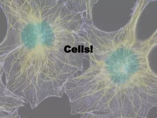
Overview: The Importance of Cells • All organisms are made of cells • The cell is the simplest collection of matter that can live • Cell structure is correlated to cellular function
LE 6-2 10 m Human height 1 m Length of some nerve and muscle cells Unaided eye 0.1 m Chicken egg 1 cm Frog egg 1 mm Measurements 1 centimeter (cm) = 10–2 meter (m) = 0.4 inch 1 millimeter (mm) = 10–3 m 1 micrometer (µm) = 10–3 mm = 10–6 m 1 nanometer (nm) = 10–3 µm = 10–9 m 100 µm Most plant and animal cells Light microscope 10 µm Nucleus Most bacteria Mitochondrion 1 µm Electron microscope Smallest bacteria 100 nm Viruses Ribosomes 10 nm Proteins Lipids 1 nm Small molecules Atoms 0.1 nm
LE 6-3a Brightfield (unstained specimen) 50 µm Brightfield (stained specimen) Phase-contrast
LE 6-3b Differential- interference- contrast (Nomarski) Fluorescence 50 µm Confocal 50 µm
Two basic types of electron microscopes (EMs) are used to study subcellular structures • Scanning electron microscopes (SEMs) focus a beam of electrons onto the surface of a specimen, providing images that look 3D • Transmission electron microscopes (TEMs) focus a beam of electrons through a specimen • TEMs are used mainly to study the internal ultrastructure of cells
LE 6-4 1 µm Scanning electron microscopy (SEM) Cilia Longitudinal section of cilium Transmission electron microscopy (TEM) Cross section of cilium 1 µm
Concept 6.2: Eukaryotic cells have internal membranes that compartmentalize their functions • The basic structural and functional unit of every organism is one of two types of cells: prokaryotic or eukaryotic • Only organisms of the domains Bacteria and Archaea consist of prokaryotic cells • Protists, fungi, animals, and plants all consist of eukaryotic cells
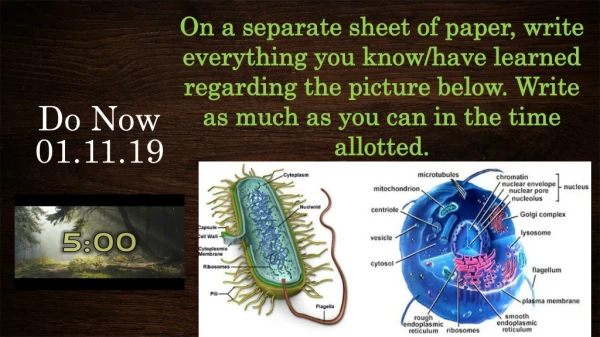
Comparing Prokaryotic and Eukaryotic Cells • Basic features of all cells: • Plasma membrane • Semifluid substance called the cytosol • Chromosomes (carry genes) • Ribosomes (make proteins)
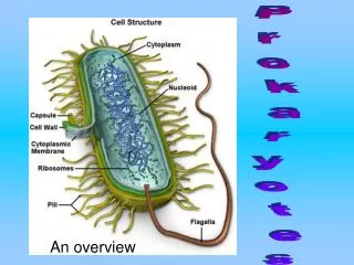
Prokaryotic cells have no nucleus • In a prokaryotic cell, DNA is in an unbound region called the nucleoid • Prokaryotic cells lack membrane-bound organelles
LE 6-6 Pili Nucleoid Ribosomes Plasma membrane Cellwall Bacterial chromosome Capsule 0.5 µm Flagella A typical rod-shaped bacterium A thin section through the bacterium Bacillus coagulans (TEM)
Eukaryotic cells have DNA in a nucleus that is bounded by a membranous nuclear envelope • Eukaryotic cells have membrane-bound organelles • Eukaryotic cells are generally much larger than prokaryotic cells • The logistics of carrying out cellular metabolism sets limits on the size of cells
LE 6-7 Surface area increases while Total volume remains constant 5 1 1 Total surface area (height x width x number of sides x number of boxes) 750 6 150 Total volume (height x width x length X number of boxes) 125 125 1 Surface-to-volume ratio (surface area volume) 6 6 1.2
The plasma membrane is a selective barrier that allows sufficient passage of oxygen, nutrients, and waste to service the volume of the cell • The general structure of a biological membrane is a double layer of phospholipids
LE 6-8 Outside of cell Carbohydrate side chain Hydrophilic region Inside of cell 0.1 µm Hydrophobic region Hydrophilic region Phospholipid Proteins Structure of the plasma membrane TEM of a plasma membrane
A Panoramic View of the Eukaryotic Cell • A eukaryotic cell has internal membranes that partition the cell into organelles • Plant and animal cells have most of the same organelles
LE 6-9a ENDOPLASMIC RETICULUM (ER Nuclear envelope Flagellum Rough ER Smooth ER NUCLEUS Nucleolus Chromatin Centrosome Plasma membrane CYTOSKELETON Microfilaments Intermediate filaments Microtubules Ribosomes: Microvilli Golgi apparatus Peroxisome Mitochondrion Lysosome In animal cells but not plant cells: Lysosomes Centrioles Flagella (in some plant sperm)
LE 6-9b Nuclear envelope Rough endoplasmic reticulum NUCLEUS Nucleolus Chromatin Smooth endoplasmic reticulum Centrosome Ribosomes (small brown dots) Central vacuole Golgi apparatus Microfilaments Intermediate filaments CYTOSKELETON Microtubules Mitochondrion Peroxisome Chloroplast Plasma membrane Cell wall Plasmodesmata Wall of adjacent cell In plant cells but not animal cells: Chloroplasts Central vacuole and tonoplast Cell wall Plasmodesmata
Concept 6.3: The eukaryotic cell’s genetic instructions are housed in the nucleus and carried out by the ribosomes • The nucleus contains most of the DNA in a eukaryotic cell • Ribosomes use the information from the DNA to make proteins
The Nucleus: Genetic Library of the Cell • The nucleus contains most of the cell’s genes and is usually the most conspicuous organelle • The nuclear envelope encloses the nucleus, separating it from the cytoplasm
LE 6-10 Nucleus Nucleus 1 µm Nucleolus Chromatin Nuclear envelope: Inner membrane Outer membrane Nuclear pore Pore complex Rough ER Surface of nuclear envelope Ribosome 1 µm 0.25 µm Close-up of nuclear envelope Pore complexes (TEM) Nuclear lamina (TEM)
Ribosomes: Protein Factories in the Cell • Ribosomes are particles made of ribosomal RNA and protein • Ribosomes carry out protein synthesis in two locations: • In the cytosol (free ribosomes) • On the outside of the endoplasmic reticulum (ER) or the nuclear envelope (bound ribosomes)
LE 6-11 Ribosomes ER Cytosol Endoplasmic reticulum (ER) Free ribosomes Bound ribosomes Large subunit Small subunit 0.5 µm TEM showing ER and ribosomes Diagram of a ribosome
The Endoplasmic Reticulum: Biosynthetic Factory • The endoplasmic reticulum (ER) accounts for more than half of the total membrane in many eukaryotic cells • The ER membrane is continuous with the nuclear envelope • There are two distinct regions of ER: • Smooth ER, which lacks ribosomes • Rough ER, with ribosomes studding its surface
Functions of Smooth ER • The smooth ER • Synthesizes lipids • Metabolizes carbohydrates • Stores calcium • Detoxifies poison
Functions of Rough ER • The rough ER • Has bound ribosomes • Produces proteins and membranes, which are distributed by transport vesicles • Is a membrane factory for the cell
The Golgi Apparatus: Shipping and Receiving Center • The Golgi apparatus consists of flattened membranous sacs called cisternae • Functions of the Golgi apparatus: • Modifies products of the ER • Manufactures certain macromolecules • Sorts and packages materials into transport vesicles
LE 6-13 Golgi apparatus cis face (“receiving” side of Golgi apparatus) Vesicles coalesce to form new cis Golgi cisternae Vesicles move from ER to Golgi 0.1 µm Vesicles also transport certain proteins back to ER Cisternae Cisternal maturation: Golgi cisternae move in a cis- to-trans direction Vesicles form and leave Golgi, carrying specific proteins to other locations or to the plasma mem- brane for secretion Vesicles transport specific proteins backward to newer Golgi cisternae trans face (“shipping” side of Golgi apparatus) TEM of Golgi apparatus
Lysosomes: Digestive Compartments • A lysosome is a membranous sac of hydrolytic enzymes • Lysosomal enzymes can hydrolyze proteins, fats, polysaccharides, and nucleic acids • Lysosomes also use enzymes to recycle organelles and macromolecules, a process called autophagy Animation: Lysosome Formation
LE 6-14a 1 µm Nucleus Lysosome Lysosome contains active hydrolytic enzymes Hydrolytic enzymes digest food particles Food vacuole fuses with lysosome Digestive enzymes Plasma membrane Lysosome Digestion Food vacuole Phagocytosis: lysosome digesting food
LE 6-14b Lysosome containing two damaged organelles 1 µm Mitochondrion fragment Peroxisome fragment Hydrolytic enzymes digest organelle components Lysosome fuses with vesicle containing damaged organelle Lysosome Digestion Vesicle containing damaged mitochondrion Autophagy: lysosome breaking down damaged organelle
Vacuoles: Diverse Maintenance Compartments • Vesicles and vacuoles (larger versions of vacuoles) are membrane-bound sacs with varied functions • A plant cell or fungal cell may have one or several vacuoles
Food vacuoles are formed by phagocytosis • Contractile vacuoles, found in many freshwater protists, pump excess water out of cells • Central vacuoles, found in many mature plant cells, hold organic compounds and water Video: Paramecium Vacuole
LE 6-15 Central vacuole Cytosol Tonoplast Central vacuole Nucleus Cell wall Chloroplast 5 µm
LE 6-16-1 Nucleus Rough ER Smooth ER Nuclear envelope
LE 6-16-2 Nucleus Rough ER Smooth ER Nuclear envelope cis Golgi Transport vesicle trans Golgi
LE 6-16-3 Nucleus Rough ER Smooth ER Nuclear envelope cis Golgi Transport vesicle Plasma membrane trans Golgi
Concept 6.5: Mitochondria and chloroplasts change energy from one form to another • Mitochondria are the sites of cellular respiration • Chloroplasts, found only in plants and algae, are the sites of photosynthesis
Chloroplasts: Capture of Light Energy • Chloroplasts contain the green pigment chlorophyll, as well as enzymes and other molecules that function in photosynthesis • Chloroplasts are found in leaves and other green organs of plants and in algae
LE 6-18 Chloroplast Ribosomes Stroma Chloroplast DNA Inner and outer membranes Granum 1 µm Thylakoid
Concept 6.6: The cytoskeleton is a network of fibers that organizes structures and activities in the cell • The cytoskeleton is a network of fibers extending throughout the cytoplasm • It organizes the cell’s structures and activities, anchoring many organelles • It is composed of three types of molecular structures: • Microtubules • Microfilaments • Intermediate filaments
LE 6-20LE 6-21a Microtubule Microfilaments 0.25 µm
Cilia and Flagella • Microtubules control the beating of cilia and flagella, locomotor appendages of some cells • Cilia and flagella differ in their beating patterns Video: Chlamydomonas Video: Paramecium Cilia
LE 6-23aLE 6-27c Direction of swimming Motion of flagella 5 µm
Cell Walls of Plants • The cell wall is an extracellular structure that distinguishes plant cells from animal cells • The cell wall protects the plant cell, maintains its shape, and prevents excessive uptake of water • Plant cell walls are made of cellulose fibers embedded in other polysaccharides and protein
LE 6-28 Central vacuole of cell Plasma membrane Secondary cell wall Primary cell wall Central vacuole of cell Middle lamella 1 µm Central vacuole Cytosol Plasma membrane Plant cell walls Plasmodesmata
The Cell: A Living Unit Greater Than the Sum of Its Parts • Cells rely on the integration of structures and organelles in order to function • For example, a macrophage’s ability to destroy bacteria involves the whole cell, coordinating components such as the cytoskeleton, lysosomes, and plasma membrane
LE 6-32 5 µm